. 2019 Nov 26;14(11):e0225851.
doi: 10.1371/journal.pone.0225851. eCollection 2019.

LFRET, a novel rapid assay for anti-tissue transglutaminase antibody detection

Affiliations

LFRET, a novel rapid assay for anti-tissue transglutaminase antibody detection

Juuso Rusanen et al. PLoS One. .

Abstract

The diagnosis of celiac disease (CD) is currently based on serology and intestinal biopsy, with detection of anti-tissue transglutaminase (tTG) IgA antibodies recommended as the first-line test. Emphasizing the increasing importance of serological testing, new guidelines and evidence suggest basing the diagnosis solely on serology without confirmatory biopsy. Enzyme immunoassays (EIAs) are the established approach for anti-tTG antibody detection, with the existing point-of-care (POC) tests lacking sensitivity and/or specificity. Improved POC methods could help reduce the underdiagnosis and diagnostic delay of CD. We have previously developed rapid homogenous immunoassays based on time-resolved Förster resonance energy transfer (TR-FRET), and demonstrated their suitability in serodiagnostics with hanta- and Zika virus infections as models. In this study, we set out to establish a protein L -based TR-FRET assay (LFRET) for the detection of anti-tTG antibodies. We studied 74 patients with biopsy-confirmed CD and 70 healthy controls, with 1) the new tTG-LFRET assay, and for reference 2) a well-established EIA and 3) an existing commercial POC test. IgG depletion was employed to differentiate between anti-tTG IgA and IgG positivity. The sensitivity and specificity of the first-generation tTG-LFRET POC assay in detection of CD were 87.8% and 94.3%, respectively, in line with those of the reference POC test. The sensitivity and specificity of EIA were 95.9% and 91.9%, respectively. This study demonstrates the applicability of LFRET to serological diagnosis of autoimmune diseases in general and of CD in particular.


Conflict of interest statement

Some of the authors are inventors in a patent "Protein L based bioassay method for determining presence of soluble antibodies in a sample and kit therefore (WO2015128548)" owned by University of Helsinki describing the LFRET assay utilized in the manuscript. (https://patentscope.wipo.int/search/en/detail.jsf?docId=WO2015128548). This does not alter our adherence to PLOS ONE policies on sharing data and materials and all authors declare that the work was carried out in accordance with good research ethics.

Figures
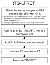
Fig 1
Fig 1. Simplified protocol for tissue transglutaminase protein L TR-FRET assay.
Eu-tTG = Europium-labeled tissue transglutaminase, AF-L = Alexa Fluor 647 -labeled protein L; TR-FRET = time-resolved Förster resonance energy transfer; RT = room temperature. We used TBS-BSA (50mM Tris-HCl, 150mM NaCl, pH 7.4, 0.2% BSA) for all component dilutions. On-plate serum dilution was 1/100 and reagent concentrations were 250 nM for AF-L and 5 nM for Eu-tTG. For further details, see the previous publication [18].
Fig 2
Fig 2. Anti-tTG-IgA FEIA results (x-axis) compared to LFRET results (y-axis) without IgG depletion.
FEIA = fluorescent enzyme immunoassay. LFRET = protein L–based time-resolved Förster resonance energy transfer assay. FEIA result is expressed as U/ml. LFRET result is expressed as average of normalized acceptor wavelength emission counts from two replicates of the same sample, with two consecutive measurements from both replicates. Patients with biopsy-confirmed celiac disease (CD) are marked with a darker spot. The solid lines indicate cutoffs for FEIA and LFRET, with the area between 7 and 10 U/ml corresponding to an equivocal result for FEIA. FEIA cutoffs are set as determined by the manufacturer. LFRET cutoffs were determined by measuring the tTG-LFRET signals for 67 tTG-antibody negative samples (as defined by FEIA) and setting the LFRET cutoff at the mean LFRET signal plus two standard deviations (SD) (35.438 + 2 × 5.316 = 46.07 counts). Pearson correlation coefficient between FEIA and LFRET results is 0.85.
Fig 3
Fig 3. Anti-tTG-IgA FEIA results (x-axis) compared to LFRET results (y-axis) with IgG depletion.
FEIA = fluorescent enzyme immunoassay. LFRET = protein L–based time-resolved Förster resonance energy transfer assay. FEIA result is expressed as U/ml. LFRET result is expressed as average of normalized acceptor wavelength emission counts from two replicates of the same sample, with two consecutive measurements from both replicates. Patients with biopsy-confirmed celiac disease (CD) are marked with a darker spot. The solid lines indicate cutoffs for FEIA and LFRET, with the area between 7 and 10 U/ml corresponding to an equivocal result for FEIA. FEIA cutoffs are set as determined by the manufacturer. LFRET cutoffs were determined by measuring the tTG-LFRET signals for 67 tTG-antibody negative samples (as defined by FEIA) and setting the LFRET cutoff at the mean LFRET signal plus two standard deviations (SD) (34.392 + 2 × 5.117 = 44.63 counts). The dashed line represents x10 upper limit of normal (ULN) for FEIA, and for LFRET a cutoff for detection of samples with a FEIA result above x10 ULN. Pearson correlation coefficient between FEIA and LFRET (with IgG depletion) results is 0.83.
Fig 4
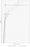
Fig 4. tTG-LFRET signal change with IgG depletion.
tTG-LFRET = tissue transglutaminase protein L–based time-resolved Förster resonance energy transfer assay. On the x-axis, x marks the LFRET signal without IgG depletion (by GullSORB treatment) and o marks the signal with IgG depletion. On the y-axis are all the studied samples, labels indicating the sample id. An algorithm for differentiation of IgG+/IgA- samples from IgA+ samples was defined as follows: First, the reduction in tTG-LFRET signal (in %) due to IgG depletion was calculated for each of the 144 study samples. The average reduction plus 2.5 × SD, corresponding to a 59% reduction in tTG-LFRET signal, was chosen as cutoff, with greater reduction taken as indication of IgG-class LFRET positivity. Second, to consider the samples IgA-negative, IgG depletion had to reduce the signal below the cutoff as determined by the mean LFRET signal plus two standard deviations (SD) (34.392 + 2 × 5.117 = 44.63 counts) of 67 tTG-antibody negative samples (as defined by FEIA). For the two anti-tTG IgA-/IgG+ samples (marked with arrows), IgG depletion reduces the LFRET signal by 1) more than 59% and 2) below the positivity cutoff. For the IgA-positive and -negative samples (all samples not marked with arrows), a similar phenomenon is not seen.

References

    1. Rubio-Tapia A, Hill ID, Kelly CP, Calderwood AH, Murray JA. ACG Clinical Guidelines: Diagnosis and Management of Celiac Disease. Am. J. Gastroenterol. 2013;108: 656–676. 10.1038/ajg.2013.79 - DOI - PMC - PubMed
    1. Ludvigsson JF, Bai JC, Biagi F, Card TR, Ciacci C, Ciclitira PJ, et al. Diagnosis and management of adult coeliac disease: guidelines from the British Society of Gastroenterology. Gut. 2014;63: 1210–1228. 10.1136/gutjnl-2013-306578 - DOI - PMC - PubMed
    1. Downey L, Houten R, Murch S, Longson D. Recognition, assessment, and management of coeliac disease: summary of updated NICE guidance. Br. Med. J. 2015;351: h4513 10.1136/bmj.h4513 - DOI - PubMed
    1. Husby S, Koletzko S, Korponay-Szabo IR, Mearin ML, Phillips A, Shamir R, et al. European Society for Pediatric Gastroenterology, Hepatology, and Nutrition guidelines for the diagnosis of coeliac disease. J. Pediatr. Gastroenterol. Nutr. 2012;54: 136–160. 10.1097/MPG.0b013e31821a23d0 - DOI - PubMed
    1. Lebwohl B, Sanders DS, Green PHR. Coeliac disease. 2018;391: 70–81. 10.1016/S0140-6736(17)31796-8 - DOI - PubMed

Publication types

MeSH terms