

Retinoic Acid and Germ Cell Development in the Ovary and Testis
- PMID: 31771306
- PMCID: PMC6995559
- DOI: 10.3390/biom9120775
Retinoic Acid and Germ Cell Development in the Ovary and Testis
Abstract
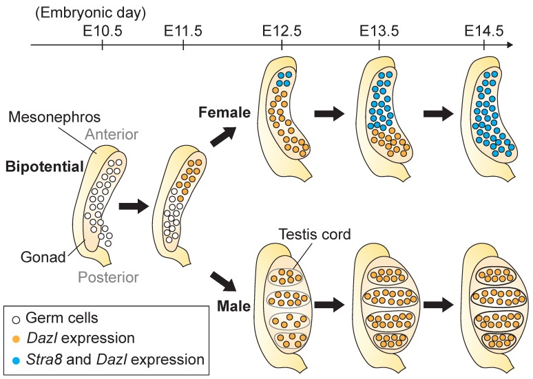
Retinoic acid (RA), a derivative of vitamin A, is critical for the production of oocytes and sperm in mammals. These gametes derive from primordial germ cells, which colonize the nascent gonad, and later undertake sexual differentiation to produce oocytes or sperm. During fetal development, germ cells in the ovary initiate meiosis in response to RA, whereas those in the testis do not yet initiate meiosis, as they are insulated from RA, and undergo cell cycle arrest. After birth, male germ cells resume proliferation and undergo a transition to spermatogonia, which are destined to develop into haploid spermatozoa via spermatogenesis. Recent findings indicate that RA levels change periodically in adult testes to direct not only meiotic initiation, but also other key developmental transitions to ensure that spermatogenesis is precisely organized for the prodigious output of sperm. This review focuses on how female and male germ cells develop in the ovary and testis, respectively, and the role of RA in this process.
Keywords: germ cells; meiosis; ovary; retinoic acid; spermatogenesis; testis.
Conflict of interest statement
The authors declare no conflict of interest.
Figures


Similar articles
-
Retinoic Acid signalling and the control of meiotic entry in the human fetal gonad.PLoS One. 2011;6(6):e20249. doi: 10.1371/journal.pone.0020249. Epub 2011 Jun 3. PLoS One. 2011. PMID: 21674038 Free PMC article.
-
Periodic production of retinoic acid by meiotic and somatic cells coordinates four transitions in mouse spermatogenesis.Proc Natl Acad Sci U S A. 2017 Nov 21;114(47):E10132-E10141. doi: 10.1073/pnas.1710837114. Epub 2017 Nov 6. Proc Natl Acad Sci U S A. 2017. PMID: 29109271 Free PMC article.
-
Retinoic acid homeostasis regulates meiotic entry in developing anuran gonads and in Bidder's organ through Raldh2 and Cyp26b1 proteins.Mech Dev. 2013 Nov-Dec;130(11-12):613-27. doi: 10.1016/j.mod.2013.09.001. Epub 2013 Sep 19. Mech Dev. 2013. PMID: 24056063
-
Roles of Retinoic Acid in Germ Cell Differentiation.Curr Top Dev Biol. 2017;125:191-225. doi: 10.1016/bs.ctdb.2016.11.013. Epub 2017 Jan 16. Curr Top Dev Biol. 2017. PMID: 28527572 Review.
-
Sex determination in mammalian germ cells: extrinsic versus intrinsic factors.Reproduction. 2010 Jun;139(6):943-58. doi: 10.1530/REP-10-0075. Epub 2010 Apr 15. Reproduction. 2010. PMID: 20395427 Review.
Cited by
-
The Molecular Signature of Human Testicular Peritubular Cells Revealed by Single-Cell Analysis.Cells. 2022 Nov 19;11(22):3685. doi: 10.3390/cells11223685. Cells. 2022. PMID: 36429113 Free PMC article.
-
Genome-wide identification, evolution and expression analysis of bone morphogenetic protein (BMP) gene family in chinese soft-shell turtle (Pelodiscus sinensis).Front Genet. 2023 Feb 1;14:1109478. doi: 10.3389/fgene.2023.1109478. eCollection 2023. Front Genet. 2023. PMID: 36816024 Free PMC article.
-
Microtubular TRIM36 E3 Ubiquitin Ligase in Embryonic Development and Spermatogenesis.Cells. 2022 Jan 12;11(2):246. doi: 10.3390/cells11020246. Cells. 2022. PMID: 35053362 Free PMC article. Review.
-
Vitamins as primary or adjunctive treatment in infertile men with varicocele: A systematic review.Arab J Urol. 2021 May 29;19(3):264-273. doi: 10.1080/2090598X.2021.1932124. eCollection 2021. Arab J Urol. 2021. PMID: 34552778 Free PMC article.
-
Advances in Female Germ Cell Induction from Pluripotent Stem Cells.Stem Cells Int. 2021 Jan 13;2021:8849230. doi: 10.1155/2021/8849230. eCollection 2021. Stem Cells Int. 2021. PMID: 33510796 Free PMC article. Review.
References
-
- Hacker A., Capel B., Goodfellow P., Lovell-Badge R. Expression of Sry, the mouse sex determining gene. Development. 1995;121:1603–1614. - PubMed
-
- Schmahl J., Eicher E.M., Washburn L.L., Capel B. Sry induces cell proliferation in the mouse gonad. Development. 2000;127:65–73. - PubMed
-
- Hilscher B., Hilscher W., Bulthoff-Ohnolz B., Kramer U., Birke A., Pelzer H., Gauss G. Kinetics of gametogenesis. I. Comparative histological and autoradiographic studies of oocytes and transitional prospermatogonia during oogenesis and prespermatogenesis. Cell Tissue Res. 1974;154:443–470. - PubMed
-
- McLaren A. Meiosis and differentiation of mouse germ cells. Symp. Soc. Exp. Biol. 1984;38:7–23. - PubMed
Publication types
MeSH terms
Substances
Grants and funding
LinkOut - more resources
Full Text Sources
