. 2019 Dec 17;116(51):26029-26037.
doi: 10.1073/pnas.1912178116. Epub 2019 Nov 26.

A critical role for microglia in maintaining vascular integrity in the hypoxic spinal cord

Affiliations

A critical role for microglia in maintaining vascular integrity in the hypoxic spinal cord

Sebok K Halder et al. Proc Natl Acad Sci U S A. .

Abstract

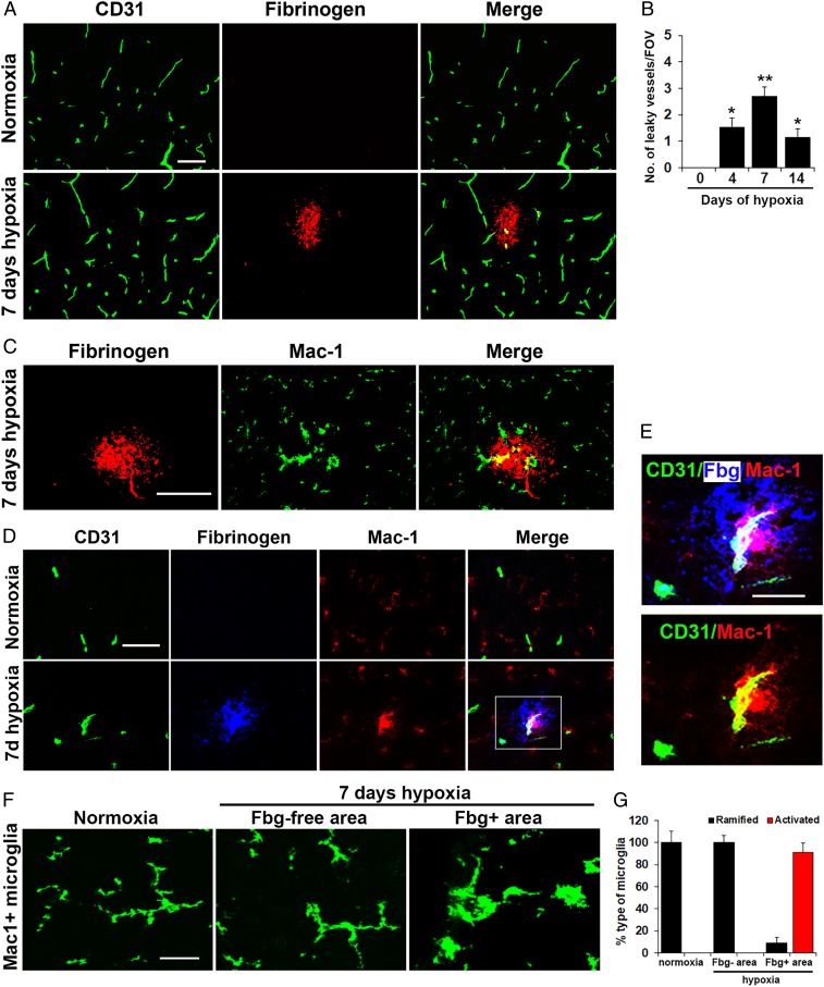
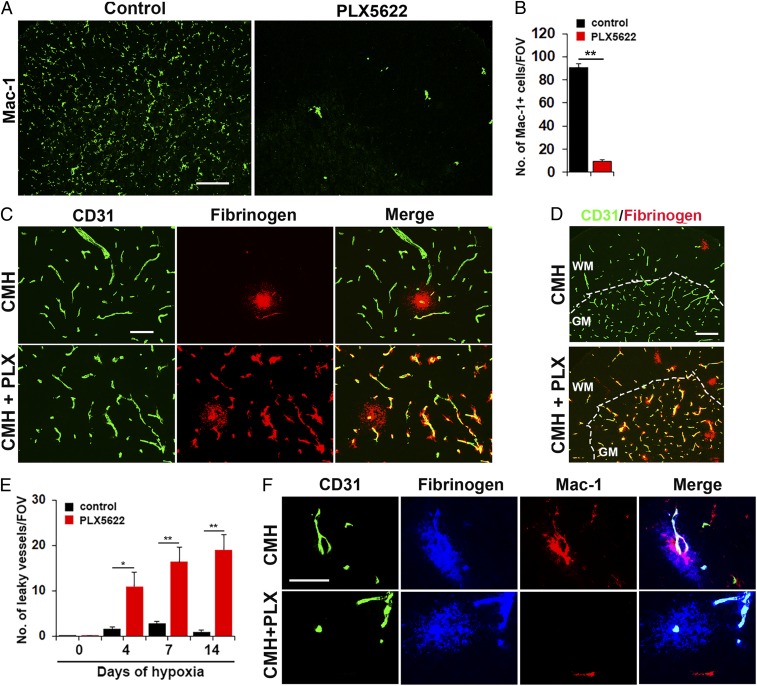
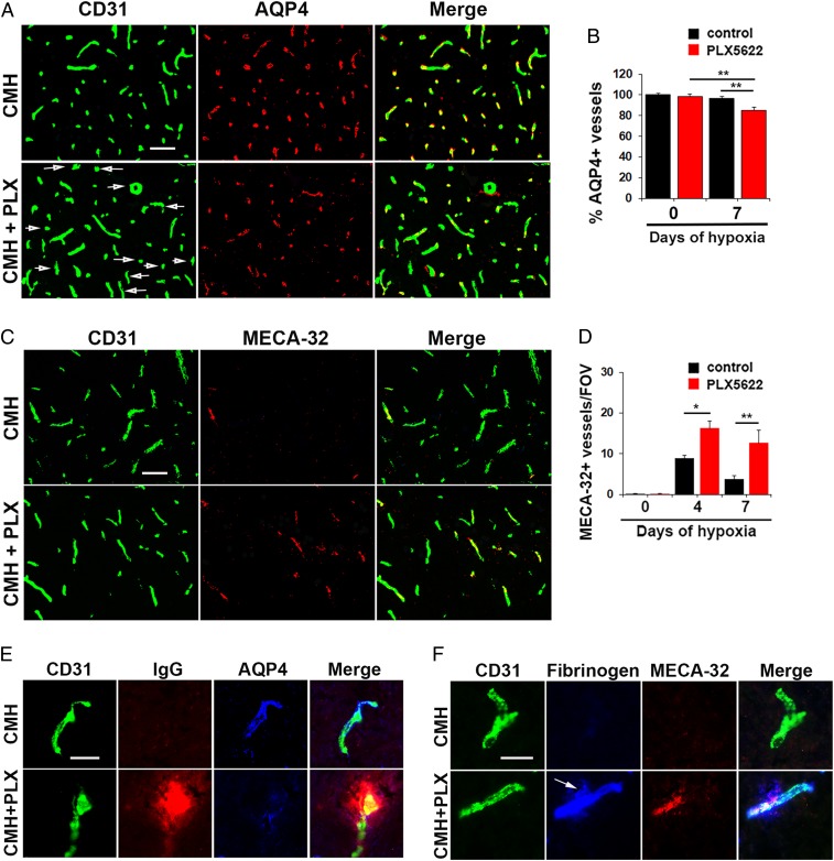
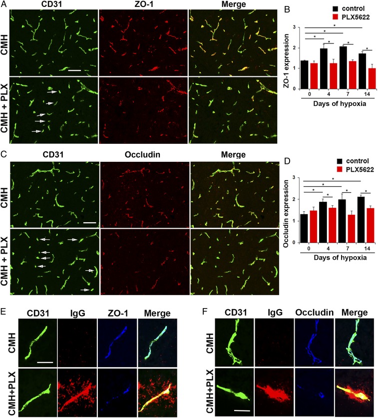
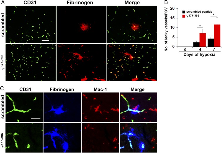
Hypoxic preconditioning reduces disease severity in a mouse model of multiple sclerosis (MS), in part by enhancing the barrier properties of spinal cord blood vessels. Because other studies have shown that similar levels of hypoxia transiently increase permeability of central nervous system (CNS) blood vessels, the goal of this study was to define the impact of chronic mild hypoxia (CMH, 8% O2) on the integrity of spinal cord blood vessels and the responses of neighboring glial cells. Using extravascular fibrinogen as a marker of vascular disruption, we found that CMH triggered transient vascular leak in spinal cord blood vessels, particularly in white matter, which was associated with clustering and activation of Mac-1-positive microglia around disrupted vessels. Microglial depletion with the colony stimulating factor-1 receptor (CSF-1R) inhibitor PLX5622, while having no effect under normoxic conditions, profoundly increased vascular leak in both white and gray matter during CMH, and this was associated with disruption of astrocyte-vascular coupling and enhanced loss of tight junction proteins. Microglial repair of leaky blood vessels was blocked by a peptide that inhibits the interaction between fibrinogen and its Mac-1 integrin receptor. These findings highlight an important role for microglia in maintaining vascular integrity in the hypoxic spinal cord and suggest that a fibrinogen-Mac-1 interaction underpins this response. As relative hypoxia is experienced in many situations including high altitude, lung disease, obstructive sleep apnea, and age-related CNS ischemia/hypoxia, our findings have important implications regarding the critical role of microglia in maintaining vascular integrity in the CNS.

Keywords: blood vessels; fibrinogen; hypoxia; microglia; spinal cord.


Conflict of interest statement

The authors declare no competing interest.

Figures

Fig. 1.
Fig. 1.
Mild hypoxic stress triggers vascular leak in spinal cord blood vessels associated with microglial clustering. Frozen sections of lumbar spinal cord taken from mice exposed to normoxia or 7-d hypoxia (8% O2) were stained for the following markers: the endothelial marker CD31 (Alexa Fluor 488) and fibrinogen (Cy-3) (A); fibrinogen (Cy-3) and Mac-1 (Alexa Fluor 488) (C); CD31 (Alexa Fluor 488), fibrinogen (abbreviated to Fbg in E) Cy-5 (blue) and Mac-1 (Cy-3) (D and E); and Mac-1 (Alexa Fluor 488) (F). (B) Quantification of the number of leaky (fibrinogen-positive) vessels/FOV. (G) Quantification of the morphological categorization of microglia under different conditions. Results are expressed as the mean ± SEM (n = 6 mice per group). *P < 0.05, **P < 0.01 vs. normoxic conditions. One-way ANOVA followed by Tukey’s multiple comparison test. Note that CMH induced transient vascular leak in spinal cord blood vessels that was associated with wrapping of Mac-1–positive microglial processes around the damaged vessel (high power images in E) and with morphological switch from ramified to activated morphology (F). (Scale bars, 50 μm; except for E, 25 μm.)
Fig. 2.
Fig. 2.
Microglial depletion results in exaggerated vascular leak during CMH. (A) Frozen sections of lumbar spinal cord taken from mice fed normal chow or PLX5622-containing chow and maintained under normoxic conditions for 7 d were stained for the microglial marker Mac-1. (B) Quantification of microglial depletion after 7-d PLX5622. Results are expressed as the mean ± SEM (n = 6 mice per group). Note that 7-d PLX5622 reduced the number of spinal cord microglia to 10% of untreated controls. (C and D) Frozen sections of lumbar spinal cord taken from mice fed normal chow or PLX5622-containing chow and maintained under hypoxic conditions for 7 d were stained for CD31 (Alexa Fluor 488) and fibrinogen (Cy-3). (E) Quantification of the number of leaky vessels/FOV. Results are expressed as the mean ± SEM (n = 6 mice per group). *P < 0.05, **P < 0.01. One-way ANOVA followed by Tukey’s multiple comparison test. Note that PLX5622-treated mice showed a much greater number of leaky blood vessels. (F) CD31/fibrinogen/Mac-1 triple-IF of control chow mice confirmed microglial clustering and elevated levels of Mac-1 expression by microglia surrounding the leaky vessel, but absence of microglial clustering in PLX5622-fed mice. (Scale bars, A, 100 μm; C, 50 μm; D, 100 μm; F, 25 μm.)
Fig. 3.
Fig. 3.
Under hypoxic conditions, absence of microglia disrupts astrocyte-vascular coupling, leading to increased MECA-32 expression. (A and C) Frozen sections of lumbar spinal cord taken from mice fed normal chow or PLX5622-containing chow and maintained under hypoxic conditions for 7 d were stained for CD31 (Alexa Fluor 488) and AQP4 (Cy-3) in A or CD31 (Alexa Fluor 488) and MECA-32 (Cy-3) in C. (B and D) Quantification of the percent of blood vessels expressing AQP4 (B) or number of blood vessels expressing MECA-32/FOV (D). Results are expressed as the mean ± SEM (n = 6 mice per group). *P < 0.05, **P < 0.01. One-way ANOVA followed by Tukey’s multiple comparison test. Note that under hypoxic conditions, spinal cords of PLX5622-fed mice contained a significant number of blood vessels that lacked AQP4 expression (see arrows) and showed greater induction of MECA-32. (E and F) High-magnification CD31/IgG/AQP4 or CD31/fibrinogen/MECA-32 triple-IF images show that under hypoxic conditions, spinal cord blood vessels in PLX5622-treated mice showed increased extravascular leak that was strongly associated with loss of AQP4 and induction of MECA-32 expression (see arrow). (Scale bars, A and C, 50 μm; E and F, 25 μm.)
Fig. 4.
Fig. 4.
Microglial depletion results in loss of endothelial tight junction protein expression during CMH. (A and C) Frozen sections of lumbar spinal cord taken from mice fed normal chow or PLX5622-containing chow and maintained under hypoxic conditions for 7 d were stained for CD31 (Alexa Fluor 488) and ZO-1 (Cy-3) in A or CD31 (Alexa Fluor 488) and occludin (Cy-3) in C. (B and D) Quantification of endothelial expression of ZO-1 (B) or occludin (D). Results are expressed as the mean ± SEM (n = 6 mice per group). *P < 0.05. One-way ANOVA followed by Tukey’s multiple comparison test. Note that under hypoxic conditions, spinal cords of PLX5622-fed mice showed focal areas in which blood vessels showed diminished expression of ZO-1 and occludin (see arrows in A and C). (E and F) High-magnification CD31/IgG/ZO-1 or CD31/IgG/occludin triple-IF images show that under hypoxic conditions, in mice fed normal chow, blood vessels expressed high levels of ZO-1 and occludin, correlating with no IgG extravascular leak, but in contrast, vessels in PLX5622-treated mice showed extravascular IgG leak that was strongly associated with disrupted endothelial ZO-1 and occludin expression. (Scale bars, A and C, 50 μm; E and F, 25 μm.)
Fig. 5.
Fig. 5.
Microglial vascular repair is blocked by a fibrinogen-derived inhibitory peptide. (A and C) Frozen sections of lumbar spinal cord taken from mice maintained under hypoxic conditions for 7 d that received daily injections of either the fibrinogen-derived inhibitory peptide γ377-395 or a scrambled peptide, were stained for CD31 (Alexa Fluor 488) and fibrinogen (Cy-3) in A or CD31 (Alexa Fluor 488), fibrinogen (Cy-5, blue), and Mac-1 (Cy-3) in C. (B) Quantification of the number of leaky vessels/FOV. Results are expressed as the mean ± SEM (n = 6 mice per group). *P < 0.05. One-way ANOVA followed by Tukey’s multiple comparison test. Note that mice treated with the inhibitory peptide γ377-395 showed a greater number of leaky blood vessels and the peptide prevented the migration and accumulation of activated microglia around leaky blood vessels (C). (Scale bars, A, 50 μm; C, 25 μm.)

References

    1. Ballabh P., Braun A., Nedergaard M., The blood-brain barrier: An overview: Structure, regulation, and clinical implications. Neurobiol. Dis. 16, 1–13 (2004). - PubMed
    1. Huber J. D., Egleton R. D., Davis T. P., Molecular physiology and pathophysiology of tight junctions in the blood-brain barrier. Trends Neurosci. 24, 719–725 (2001). - PubMed
    1. Pardridge W. M., Blood-brain barrier drug targeting: The future of brain drug development. Mol. Interv. 3, 90–105 (2003). - PubMed
    1. Daneman R., Zhou L., Kebede A. A., Barres B. A., Pericytes are required for blood-brain barrier integrity during embryogenesis. Nature 468, 562–566 (2010). - PMC - PubMed
    1. del Zoppo G. J., Milner R., Integrin-matrix interactions in the cerebral microvasculature. Arterioscler. Thromb. Vasc. Biol. 26, 1966–1975 (2006). - PubMed

Publication types

MeSH terms