. 2019 Dec 17;116(51):25745-25755.
doi: 10.1073/pnas.1906117116. Epub 2019 Nov 26.

Solenodon genome reveals convergent evolution of venom in eulipotyphlan mammals

Affiliations

Solenodon genome reveals convergent evolution of venom in eulipotyphlan mammals

Nicholas R Casewell et al. Proc Natl Acad Sci U S A. .

Abstract

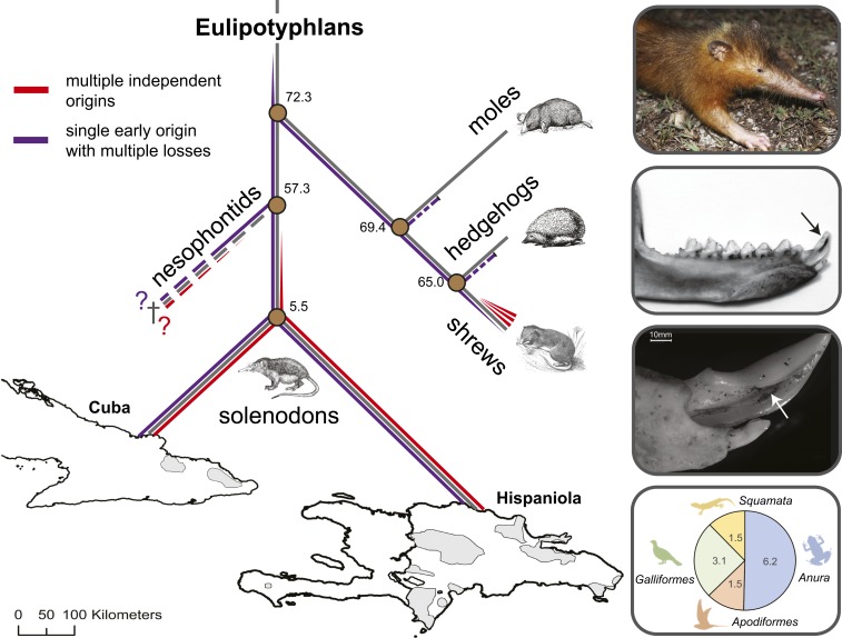
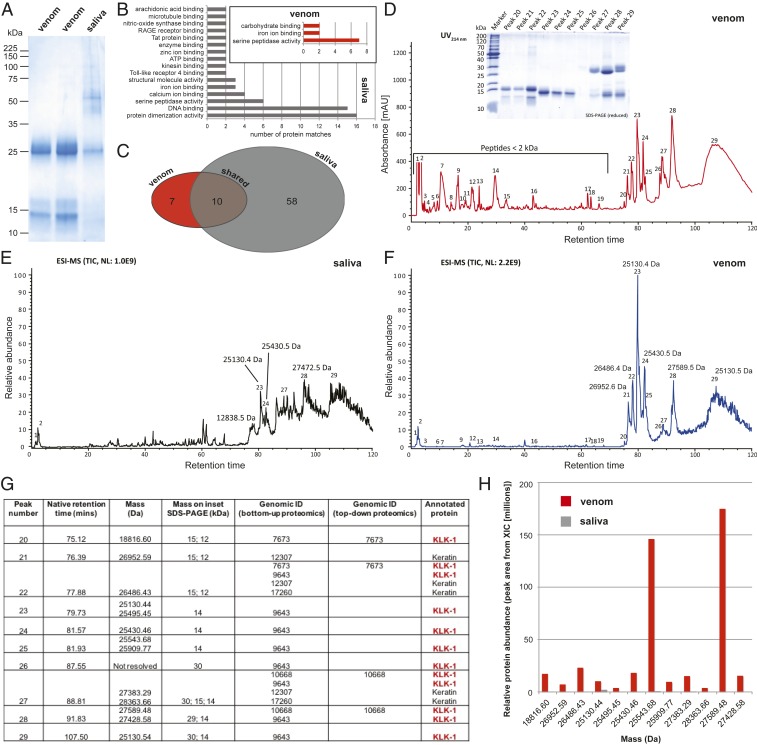
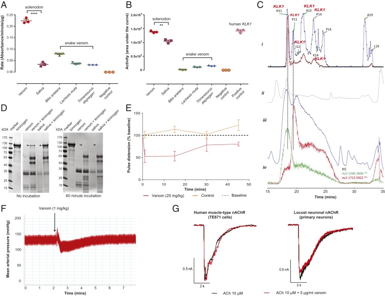
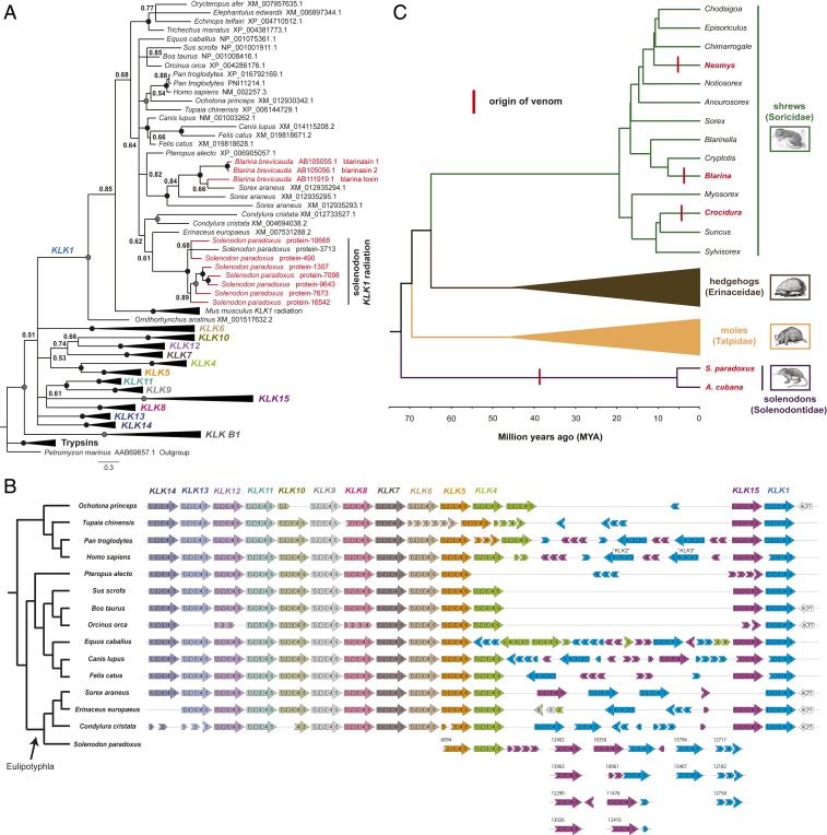
Venom systems are key adaptations that have evolved throughout the tree of life and typically facilitate predation or defense. Despite venoms being model systems for studying a variety of evolutionary and physiological processes, many taxonomic groups remain understudied, including venomous mammals. Within the order Eulipotyphla, multiple shrew species and solenodons have oral venom systems. Despite morphological variation of their delivery systems, it remains unclear whether venom represents the ancestral state in this group or is the result of multiple independent origins. We investigated the origin and evolution of venom in eulipotyphlans by characterizing the venom system of the endangered Hispaniolan solenodon (Solenodon paradoxus). We constructed a genome to underpin proteomic identifications of solenodon venom toxins, before undertaking evolutionary analyses of those constituents, and functional assessments of the secreted venom. Our findings show that solenodon venom consists of multiple paralogous kallikrein 1 (KLK1) serine proteases, which cause hypotensive effects in vivo, and seem likely to have evolved to facilitate vertebrate prey capture. Comparative analyses provide convincing evidence that the oral venom systems of solenodons and shrews have evolved convergently, with the 4 independent origins of venom in eulipotyphlans outnumbering all other venom origins in mammals. We find that KLK1s have been independently coopted into the venom of shrews and solenodons following their divergence during the late Cretaceous, suggesting that evolutionary constraints may be acting on these genes. Consequently, our findings represent a striking example of convergent molecular evolution and demonstrate that distinct structural backgrounds can yield equivalent functions.

Keywords: convergent molecular evolution; gene duplication; genotype phenotype; kallikrein toxin; venom systems.


Conflict of interest statement

The authors declare no competing interest.

Figures

Fig. 1.
Fig. 1.
The 2 competing hypotheses relating to the origin of venom in eulipotyphlans and key characteristics of the Hispaniolan solenodon (S. paradoxus). The schematic phylogeny (gray lines) highlights the estimated divergence times of eulipotyphlan families and solenodon species and the 2 competing hypotheses relating to the origin of venom in this group. Purple lines indicate the early origin of venom hypothesis followed by losses in moles, hedgehogs, and some shrews (narrowing line), whereas red lines indicate the alternative hypothesis of multiple independent origins of venom in shrews (3 times), solenodons, and possibly nesophontids. Shaded areas of the map indicate the modern distribution of the 2 solenodon species (A. cubana and S. paradoxus) on the islands of Cuba and Hispaniola, respectively. Boxes on the Right show (from Top to Bottom): a wild specimen of S. paradoxus, its lower jaw morphology, its enlarged tubular lower second incisor used for venom delivery visualized via stereo microscopy, and the composition and frequency of occurrence (in percentages) of vertebrates detected in their diet determined by DNA barcoding analyses of fecal samples. Divergence times displayed on the phylogeny are from refs. and . The photograph of the wild solenodon is courtesy of Rocio Pozo.
Fig. 2.
Fig. 2.
Proteomic analyses of Hispaniolan solenodon (S. paradoxus) venom and saliva reveal KLK1 proteins as major venom components. (A) Reduced SDS-PAGE gel electrophoretic profiles of venom and saliva samples. (B) Gene ontology (GO) term analysis of proteins identified via shotgun proteomic-based annotation to the genome. GO term categories are only displayed for those with at least 2 matches. (C) Venn diagram displaying the number of proteins in the venom and saliva, and those identified in both samples via shotgun proteomic-based annotation to the genome. (D) Reverse-phase chromatographic separation of venom. Venom was separated by semipreparative reversed-phase HPLC (UV214nm) and manually collected. Peptides were directly submitted to LC-MS/MS, whereas protein fractions were analyzed by SDS-PAGE (Inset) under reducing conditions. Afterward, protein bands were subjected to in-gel trypsin digestion and identified by spectrum peptide matching against the translated S. paradoxus genome database. (E and F) LC-top-down MS analysis of saliva (E) and venom (F). The peak nomenclature is based on the chromatogram fractions, shown in D. (E) Total ion current (TIC) profile of native saliva separated by HPLC. (F) TIC profile of native venom separated by HPLC. (G) Summary table of the proteins identified via top-down and bottom-up proteomic analyses of solenodon venom, including their mass, corresponding identification in the genome (genome ID), and protein annotation. All identified proteins are annotated as KLK1, with the exception of keratins, which are human contaminants. (H) Comparison of the relative abundance of main proteins present in chromatographic fractions of venom and saliva from top-down MS experiments. SI Appendix, Table S2 presents a summary of the protein matches identified in solenodon venom and saliva via the various proteomic approaches.
Fig. 3.
Fig. 3.
Functional assessments of Hispaniolan solenodon (S. paradoxus) venom reveals kallikrein serine protease activity and hypotensive effects. Solenodon venom has extensive (A) serine protease activity, as measured by chromogenic enzyme assay, and (B) plasminogen-activating activity, as measured by fluorescent enzyme assay. The data displayed are the mean rate of substrate consumption (A) or area under the kinetic curve (B) for mean measurements (±SEM) taken from 3 independent experiments; ****P < 0.0001; **P < 0.01; unpaired 2-tailed t tests. (C) Nanofractionation bioassaying reveals that KLK1 proteins are responsible for plasminogen-activating activity. (i) Bioactivity chromatogram at 5 mg/mL (blue line) and 1 mg/mL (red line) venom show the activity of each fraction, where positive peaks represent bioactive compounds. Bioactive wells selected for tryptic digestion are indicated by green arrows and well numbers, and those identified by mass spectrometry as KLK1s are labeled in red. (ii) UV trace at 254 nm collected during the LC-MS run with a UV-visible spectroscopy detector. (iii) TIC shown by the LC-MS chromatogram. (iv) Extracted ion currents (XICs) of the m/z values from the LC-MS data corresponding to the bioactives detected in the plasminogen assay. (D) Solenodon venom degrades high molecular weight kininogen more potently than saliva without incubation. SDS-PAGE gel electrophoresis profiles demonstrate that both venom and saliva completely degrade kininogen (arrows) when preincubated for 60 min, but that venom also degrades kininogen in the absence of preincubation. (E) Solenodon venom causes substantial reductions in the pulse distension of envenomed mice (25 mg/kg; n = 3) when compared to baseline measurements and controls (saline; n = 3). The data displayed represent mean measurements, and the error bars represent SDs. (F) Solenodon venom causes a transient depressor effect on the mean arterial blood pressure of the anesthetized rat. The data displayed are a representative trace from 1 of 5 experimental animals that received 1 mg/kg venom (see also SI Appendix, Fig. S3). (G) Solenodon venom has no effect on nicotinic acetylcholine receptors. Representative whole-cell patch-clamp traces showing human muscle-type TE671 (Left) and locust neuron nAChR (Right) responses to 10 µM acetylcholine and the coapplication of acetylcholine with 5 µg/mL solenodon venom. VH = −75 mV.
Fig. 4.
Fig. 4.
Molecular analyses reveal that eulipotyphlan venom systems and their toxin constituents have evolved independently by convergent evolution. (A) Molecular phylogeny of amino acid translations of tetrapod KLKs demonstrate that solenodon KLK1 venom genes form a strongly supported monophyly and are polyphyletic to Blarina shrew venom genes. The phylogeny was derived by Bayesian inference analysis (n = 106; 2 × 10⁸ generations, 4 parallel runs with 6 simultaneous MCMC simulations). Genes encoding for proteins detected in solenodon venom (SI Appendix, Table S2) or Blarina venom (11, 33) are highlighted by red-colored branches and tip labels. Support values represent Bayesian posterior probabilities (BPP), where black circles represent BPP = 1.00 and gray circles BPP ≥ 0.95. See also SI Appendix, Fig. S4 for the nucleotide-derived phylogeny. (B) Analysis of the genomic organization of mammalian KLKs demonstrates that KLK1s are atypically numerous in the solenodon. Distinct patterns of KLK1 orientation across eulipotyphlans suggest that venom genes have arisen independently in the solenodon, and evidence of multiple solenodon genome scaffolds containing KLK1 and KLK15 adjacent to one another suggests that these may form the basis of a duplication cassette. (C) Ancestral state reconstruction of the origin of venom in eulipotyphlans reveals that venom most likely evolved independently on 4 occasions (red vertical lines). Genera containing venomous species (or the species themselves) are highlighted by red tip labels. The computed ancestral traits for each node are depicted by pie charts, where the proportion of red color represents the posterior probability of the most recent common ancestor being venomous, and blue represents nonvenomous. In all cases, ancestral nodes support the nonvenomous character state with a posterior probability of 1.00, except for the Suncus and Crocidura node, where the support value was greater than 0.85. Divergence times are indicated by the scale, and these, along with the tree topology, are derived from prior studies (15, 19, 37). The specific timing of the origin of venom should not be inferred from the placement of the vertical red bars on the tree—these are placed arbitrarily at the midpoint of each relevant branch.

Similar articles

Cited by

References

    1. Casewell N. R., Wüster W., Vonk F. J., Harrison R. A., Fry B. G., Complex cocktails: The evolutionary novelty of venoms. Trends Ecol. Evol. 28, 219–229 (2013). - PubMed
    1. Fry B. G., et al. , The toxicogenomic multiverse: Convergent recruitment of proteins into animal venoms. Annu. Rev. Genomics Hum. Genet. 10, 483–511 (2009). - PubMed
    1. Duda T. F. Jr, Palumbi S. R., Molecular genetics of ecological diversification: Duplication and rapid evolution of toxin genes of the venomous gastropod Conus. Proc. Natl. Acad. Sci. U.S.A. 96, 6820–6823 (1999). - PMC - PubMed
    1. Dowell N. L., et al. , The deep origin and recent loss of venom toxin genes in rattlesnakes. Curr. Biol. 26, 2434–2445 (2016). - PMC - PubMed
    1. Brust A., et al. , Differential evolution and neofunctionalization of snake venom metalloprotease domains. Mol. Cell. Proteomics 12, 651–663 (2013). Correction in: Mol Cell Proteomics12, 1488 (2013). - PMC - PubMed

Publication types

LinkOut - more resources