. 2019 Nov 26;10(1):5389.
doi: 10.1038/s41467-019-12973-1.

An MPER antibody neutralizes HIV-1 using germline features shared among donors

Collaborators, Affiliations

An MPER antibody neutralizes HIV-1 using germline features shared among donors

Lei Zhang et al. Nat Commun. .

Abstract

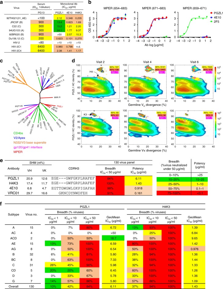
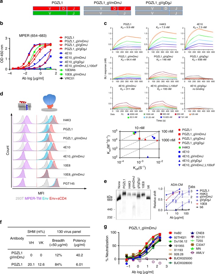
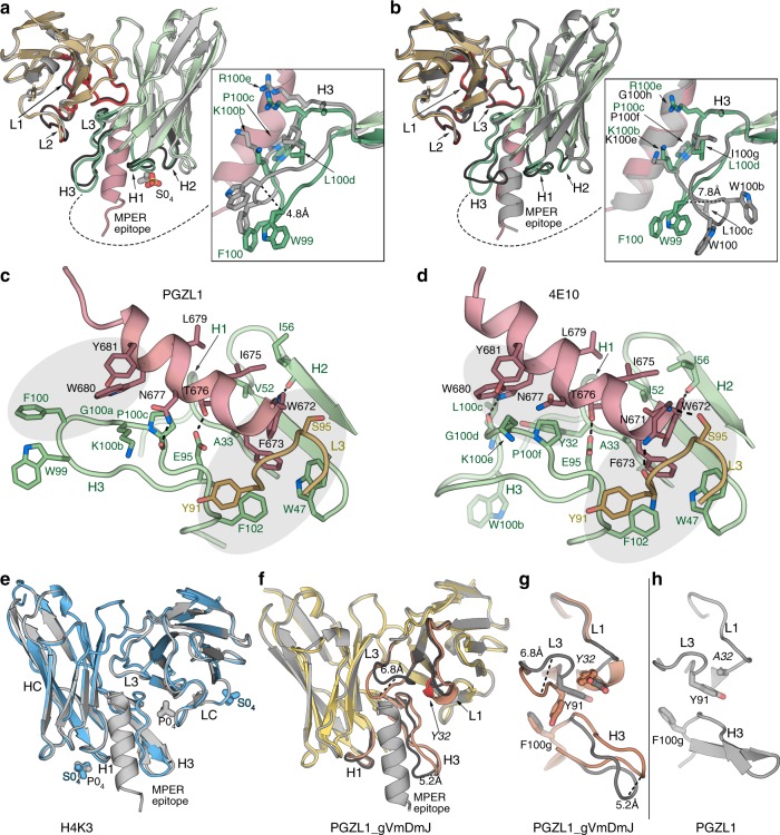
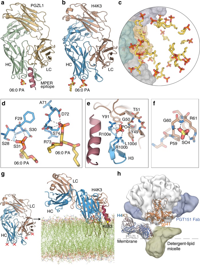
The membrane-proximal external region (MPER) of HIV-1 envelope glycoprotein (Env) can be targeted by neutralizing antibodies of exceptional breadth. MPER antibodies usually have long, hydrophobic CDRH3s, lack activity as inferred germline precursors, are often from the minor IgG3 subclass, and some are polyreactive, such as 4E10. Here we describe an MPER broadly neutralizing antibody from the major IgG1 subclass, PGZL1, which shares germline V/D-region genes with 4E10, has a shorter CDRH3, and is less polyreactive. A recombinant sublineage variant pan-neutralizes a 130-isolate panel at 1.4 μg/ml (IC50). Notably, a germline revertant with mature CDR3s neutralizes 12% of viruses and still binds MPER after DJ reversion. Crystal structures of lipid-bound PGZL1 variants and cryo-EM reconstruction of an Env-PGZL1 complex reveal how these antibodies recognize MPER and viral membrane. Discovery of common genetic and structural elements among MPER antibodies from different patients suggests that such antibodies could be elicited using carefully designed immunogens.


Conflict of interest statement

The authors declare the following competing interests: A U.S. provisional patent application entitled “Human broadly neutralizing antibodies against the membrane-proximal external region of HIV gp41” is to be filed covering the manuscript in its entirety, with the currently named inventors L.Z., A.I., J.Z., I.A.W., and M.B.Z., all of whom are currently employed by or affiliated with The Scripps Research Institute. All other authors declare no competing interests.

Figures

Fig. 1
Fig. 1
Properties of an MPER-targeted bnAb. a Neutralization of HIV-1 six-virus panel and HIV-2 (HIV-1 MPER) chimeras by PG13 plasma and monoclonal antibodies PGZL1, 4E10, and H4K3. b ELISA binding of PGZL1 to MPER peptides, using 4E10 and 2F5 as controls. c Maximum likelihood (ML) tree of HC variable regions of described bnAbs, colored by Env specificity. d Divergence/identity analysis of donor PG13 antibody repertoire over three visits in 9 months. NGS-derived antibody chains are plotted as a function of sequence identity to PGZL1 and divergence from their putative germline genes. Colors indicate sequence density. Sequences with a CDR3 identity of ≥80/85% (HC/KC) and with a CDR3 identity of ≥95% are shown as yellow and orange dots on the 2D plots, with the number of sequences highlighted in yellow and orange shades, respectively. Sequences bioinformatically selected for synthesis are shown as magenta stars on the 2D plots, with the number of sequences (Syn) highlighted in magenta shade. e, f Neutralization breadth and potency of PGZL1 and H4K3 against a 130-virus panel (e) and the same data as in e but subdivided by HIV subtype (f).
Fig. 2
Fig. 2
Characterization of germline-reverted antibody PGZL1 gVmDmJ. a Cartoon of mature PGZL1 VH (red; top) and Vκ (green; bottom) subdivided by V, D, and J regions, and germline reversions (gray) to create PGZL1 gVmDmJ (middle) and PGZL1 gVgDgJ (right). b ELISA binding of PGZL1 germline revertants to MPER peptide, using analogous 10E8 and 4E10 controls. c BLI-binding kinetics of PGZL1 variants to immobilized MPER peptide (top panels). 4E10 variants were also used for comparison. kon and koff of antibodies are shown on a scatter plot with affinity constant, KD, as dashed lines (bottom). d Cells expressing MPER-TM (purple histograms, left) were stained in flow cytometry by mature and germline-reverted antibodies at 2 μg/ml. HIV Env (right) in the presence and absence of soluble CD4 (sCD4; red and blue histograms, respectively) were stained by mature and germline-reverted antibodies at 2 and 10 μg/ml, respectively. e BN-PAGE Env mobility shift assay. HIV-1 virions were incubated with Fab PGZL1 and H4K3 (20 µg/ml), or PGZL1 gVmDmJ and gVgDgJ (200 µg/ml). 10E8 (20 µg/ml) and non-neutralizing antibody b6 (200 µg/ml) were used as positive and negative controls, respectively. Relative shift and stoichiometry of Fab to Env was quantified. The error bars represent the SD of n = 2 biologically independent experiments. f Neutralization potency and breadth of PGZL1 gVmDmJ against a 130-virus panel of HIV-1 in TZM-bl assay at 200 μg/ml. g Neutralization of 13 isolates sensitive to PGZL1 gVmDmJ chosen from the 130-virus panel. Source data for bg are provided as a Source Data file.
Fig. 3
Fig. 3
Dominant role of CDRH3 in PGZL1 HIV-1 neutralization by D-gene-encoded residues. a Fold decrease in neutralization (IC50) of isolate 92TH021 relative to wild-type PGZL1 by CDR grafts and LC substitution from 4E10 (left), and by 4E10 substitutions L100cF and PGZL1 LC (right). b Neutralization (log IC50) of a six-virus panel by PGZL1 and 4E10 variant antibodies. c Effect of Ala substitutions in DH-encoded residues W99 and F100 on the ability of PGZL1 mature (top panels) and inferred germline antibodies (bottom panels) to neutralize Du156.12 and HxB2 (left panels), as well as to bind MPER peptide in an ELISA (right panels). d Antibody polyreactivity in an ELISA as a function of area under the curve (AUC) of PGZL1 and 4E10 variant antibodies against nonspecific antigens. VRC01 is a negative control. Two-way ANOVA multiple comparisons was used to compare the difference between groups (n = 7, **p = 0.0062, ***p = 0.0001, ****p < 0.0001). e Immunofluorescence staining of HEp-2 cells. Antibodies were tested at 50 μg/ml using 4E10 and VRC01 as positive and negative controls, respectively; images are at ×200 magnification and the scale bar is 400 µm. Source data for bd are provided as a Source Data file.
Fig. 4
Fig. 4
PGZL1 variant crystal structures and comparison with 4E10. a Superposition of the crystal structures of the mature PGZL1 variable domain (from the Fab) bound to MPER671-683 (wheat, LC; green, HC; pink, MPER) and unbound PGZL1 (gray). CDRs of the bound structure are shown in red (LC) and green (HC), and CDRs of the unbound structure are shown in black. Inset: superposition of free and bound CDRH3 with residues near the MPER shown as sticks. b Superposition of PGZL1–MPER671-683 and 4E10-MPER671-683 (gray; PDB 2FX7 [https://www.rcsb.org/structure/2fx7]). Coloring and inset as in a. c PGZL1–MPER671-683 combining site (wheat, LC; green, HC; pink, MPER; interacting residues - sticks). Shaded regions highlight aromatic clusters. d 4E10-MPER671-683 combining site, colored as in c. e Superposition of unbound (blue) and MPER671-683-bound (gray) H4K3. Ions are shown as sticks. f Superposition of unbound (yellow, CDR loops, brown) and MPER671-683-bound (gray) PGZL1 gVmDmJ. g CDR loops in bound (gray) and unbound (brown) PGZL1 gVmDmJ with residues that influence loop conformations shown as sticks. h Same region as in g for the mature PGZL1 structure.
Fig. 5
Fig. 5
Lipid binding and angle of approach of PGZL1 variants to the viral membrane. a Cartoon of the PGZL1-MPER671-683 complex structure (wheat, LC; green, HC; pink, MPER) crystallized with 06:0 PA (sticks). b Cartoon of H4K3 (brown, LC; blue, HC) crystallized with 06:0 PA (sticks). c Stick rendering of 06:0 PA fragments forming a lipid vesicle at the interface of 12 crystallographic and non-crystallographic-related H4K3 Fabs. The four Fabs in the asymmetric unit are shown as gray, green, yellow, and blue color surfaces. d Stick rendering of observed lipid-binding sites in H4K3. e Phosphate-binding site near to H4K3 CDRH3 when MPER671-683 (pink) is bound. Colors as in b. f Sulfate-binding site in FRL3 of H4K3. g Model of H4K3 binding to the MPER (red)-viral membrane (green) epitope. The model at the right side of the arrow was built based on the regions where experimental lipids and anions (red X) bind on H4K3 (left side of the arrow); cognate lipids are shown as sticks inside the modeled membrane. The position of the MPER K683 residue is indicated with a yellow dot. h Cryo-EM reconstruction of full-length AMC011-PGT151-PGZL1 complex at 8.9 Å with H4K3 (blue/brown ribbons) fitted into the Env density at the base of the gp41 stem. The MPER is shown as a red ribbon and lipid head groups as sticks. The detergent–lipid micelle is shown in olive and PGT151 density in blue. Dashed lines show the approximate location where the outer surface of the membrane would be on the virus or infected cells.
Fig. 6
Fig. 6
MPER-induced PO4-binding site of H4K3 and PGZL1 lipid site mutant characterization and electrostatics. a, b Stick rendering of the PO4-binding site in (a) H4K3 and (b) PGZL1. The side chain at LC position 50 in each antibody is surrounded by dots. Select HC residues of the two antibodies are shown in blue (a) and green (b), and the LC is shown in brown. MPER is shown as a pink ribbon. c Binding of H4K3 lipid-binding site mutants to immobilized MPER peptide by BLI. d Binding of H4K3 lipid-binding site mutants to MPER peptide by ELISA and neutralization (log IC50) of HxB2 and Du156.12. e BLI, ELISA binding to MPER peptide, and neutralization statistics. fi Surface rendering, along the MPER helical axis (red ribbon), of the solvent accessible electrostatic potential contoured at ± 5 kT/e for (f) PGZL1 gVmDmJ, (g) PGZL1, (h) H4K3, and (i) 4E10. Observed lipid fragments and anions are shown as sticks. Source data for h and i are provided as a Source Data file.

References

    1. Burton DR, Hangartner L. Broadly neutralizing antibodies to HIV and their role in vaccine design. Annu. Rev. Immunol. 2016;34:635–659. doi: 10.1146/annurev-immunol-041015-055515. - DOI - PMC - PubMed
    1. Kwong PD, Mascola JR. HIV-1 vaccines based on antibody identification, B cell ontogeny, and epitope structure. Immunity. 2018;48:855–871. doi: 10.1016/j.immuni.2018.04.029. - DOI - PubMed
    1. Molinos-Albert LM, Clotet B, Blanco J, Carrillo J. Immunologic insights on the membrane proximal external region: a major human immunodeficiency virus type-1 vaccine target. Front. Immunol. 2017;8:1154. doi: 10.3389/fimmu.2017.01154. - DOI - PMC - PubMed
    1. Haynes BF, Moody MA, Verkoczy L, Kelsoe G, Alam SM. Antibody polyspecificity and neutralization of HIV-1: a hypothesis. Hum. Antibodies. 2005;14:59–67. doi: 10.3233/HAB-2005-143-402. - DOI - PMC - PubMed
    1. Krebs SJ, et al. Longitudinal analysis reveals early development of three MPER-directed neutralizing antibody lineages from an HIV-1-infected individual. Immunity. 2019;50:677–691 e613. doi: 10.1016/j.immuni.2019.02.008. - DOI - PMC - PubMed

Publication types

MeSH terms