Review
. 2020 Feb;25(2):351-367.
doi: 10.1038/s41380-019-0609-8. Epub 2019 Nov 26.

Microglial regional heterogeneity and its role in the brain

Affiliations
Review

Microglial regional heterogeneity and its role in the brain

Yun-Long Tan et al. Mol Psychiatry. 2020 Feb.

Abstract

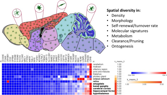
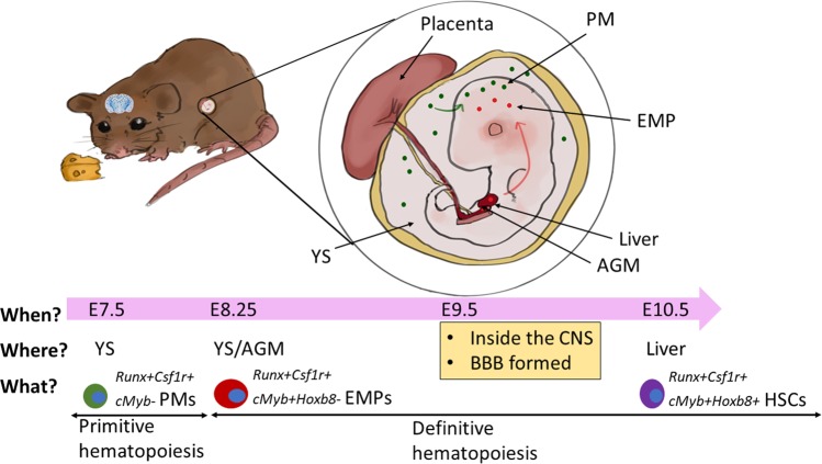
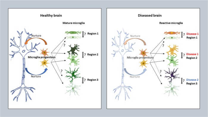
Microglia have been recently shown to manifest a very interesting phenotypical heterogeneity across different regions in the mammalian central nervous system (CNS). However, the underlying mechanism and functional meaning of this phenomenon are currently unclear. Baseline diversities of adult microglia in their cell number, cellular and subcellular structures, molecular signature as well as relevant functions have been discovered. But recent transcriptomic studies using bulk RNAseq and single-cell RNAseq have produced conflicting results on region-specific signatures of microglia. It is highly speculative whether such spatial heterogeneity contributes to varying sensitivities of individual microglia to the same physiological and pathological signals in different CNS regions, and hence underlie their functional relevance for CNS disease development. This review aims to thoroughly summarize up-to-date knowledge on this specific topic and provide some insights on the potential underlying mechanisms, starting from microgliogenesis. Understanding regional heterogeneity of microglia in the context of their diverse neighboring neurons and other glia may provide an important clue for future development of innovative therapies for neuropsychiatric disorders.


Conflict of interest statement

The authors declare that they have no conflict of interest.

Figures

Fig. 1
Fig. 1
Regional heterogeneity of microglia in the brain. Microglia differ in their cell number, cellular and subcellular structures, molecular signature as well as relevant functions in different mouse brain areas. Microgliome genes were selected based on several seminal transcriptomic studies [25, 26, 63, 68, 107, 115, 140] and their RNAseq data in 13 mouse brain regions were retrieved from the human protein atlas. An expression heatmap was drawn in Morpheus (Broad Institute). Brain regions were clustered according to K-means of expression levels (grouping score at 5, Pearson’s method). The data are also provided in Supplementary Table 1
Fig. 2
Fig. 2
Possible different regional ontogenesis and entry routes of microglia in the developing mouse brain. Microglia, along with other tissue macrophages, are generated in successive waves of myelogenesis during mouse embryonic (E) development. The first wave of microglia is generated by Runx- and Csf1r-dependent, cMyb-independent primitive macrophages (PMs) in the yolk sac (YS) at E7.5, whilst the second wave is generated by cMyb-dependent erythromyeloid progenitors (EMPs) at E8.25 from the YS as well as the aorta–gonad–mesonephros (AGM) and fetal liver at E8.25-E10, during which definitive hematopoiesis happens. Adult microglial (or microglia-like) cells may also come from cMyb-dependent fetal liver monocytes in mouse [141] and embryonic hematopoietic stem cells (HSCs) in zebrafish [94]. A transient appearance of fetal liver-derived microglia in the neonate mouse brain was also found [46]. Microglial precursors from the primitive and definitive waves migrate into the brain at E9.5 and self-sustain throughout adulthood [91, 92]. It is possible that microglia deriving from different origins and in different waves may enter and occupy different niches in the developing brain. For example, Hoxb8 and Hoxb+ microglia came from primitive and definitive hematopoiesis, respectively, and infiltrated the brain at different E stages in different brain areas [101]
Fig. 3
Fig. 3
Can regional heterogeneity of microglia be plastic and dynamic under healthy and diseased conditions? In the healthy adult CNS, a balanced neuron-microglia interaction may be necessary, or unnecessary, for microglia derived from progenitors of singular or multiple lineages to maturate and maintain their individual identities in different regions. Whether their phenotypes and the related functions are interchangeable under normal condition is unclear. Under pathological conditions, microglia at different locations, or even in the same place, probably do not all react homogenously toward different stimuli at different places and may individually change features during the process of the disease development. But whether they can dynamically transform into another subtype within the same location or even into a subtype that may carry different anatomical features is also uncertain

References

    1. Zeisel A, Munoz-Manchado AB, Codeluppi S, Lonnerberg P, La Manno G, Jureus A, et al. Brain structure. Cell types in the mouse cortex and hippocampus revealed by single-cell RNA-seq. Science. 2015;347:1138–42. doi: 10.1126/science.aaa1934. - DOI - PubMed
    1. Tasic B, Menon V, Nguyen TN, Kim TK, Jarsky T, Yao Z, et al. Adult mouse cortical cell taxonomy revealed by single cell transcriptomics. Nat Neurosci. 2016;19:335–46. doi: 10.1038/nn.4216. - DOI - PMC - PubMed
    1. Ko Y, Ament SA, Eddy JA, Caballero J, Earls JC, Hood L, et al. Cell type-specific genes show striking and distinct patterns of spatial expression in the mouse brain. Proc Natl Acad Sci USA. 2013;110:3095–3100. doi: 10.1073/pnas.1222897110. - DOI - PMC - PubMed
    1. Marques S, Zeisel A, Codeluppi S, van Bruggen D, Mendanha Falcao A, Xiao L, et al. Oligodendrocyte heterogeneity in the mouse juvenile and adult central nervous system. Science. 2016;352:1326–9. doi: 10.1126/science.aaf6463. - DOI - PMC - PubMed
    1. John Lin CC, Yu K, Hatcher A, Huang TW, Lee HK, Carlson J, et al. Identification of diverse astrocyte populations and their malignant analogs. Nat Neurosci. 2017;20:396–405. doi: 10.1038/nn.4493. - DOI - PMC - PubMed

Publication types