. 2019 Oct 23:2019:6708435.
doi: 10.1155/2019/6708435. eCollection 2019.

Combining ECM Hydrogels of Cardiac Bioactivity with Stem Cells of High Cardiomyogenic Potential for Myocardial Repair

Affiliations

Combining ECM Hydrogels of Cardiac Bioactivity with Stem Cells of High Cardiomyogenic Potential for Myocardial Repair

Rui Bai et al. Stem Cells Int. .

Abstract

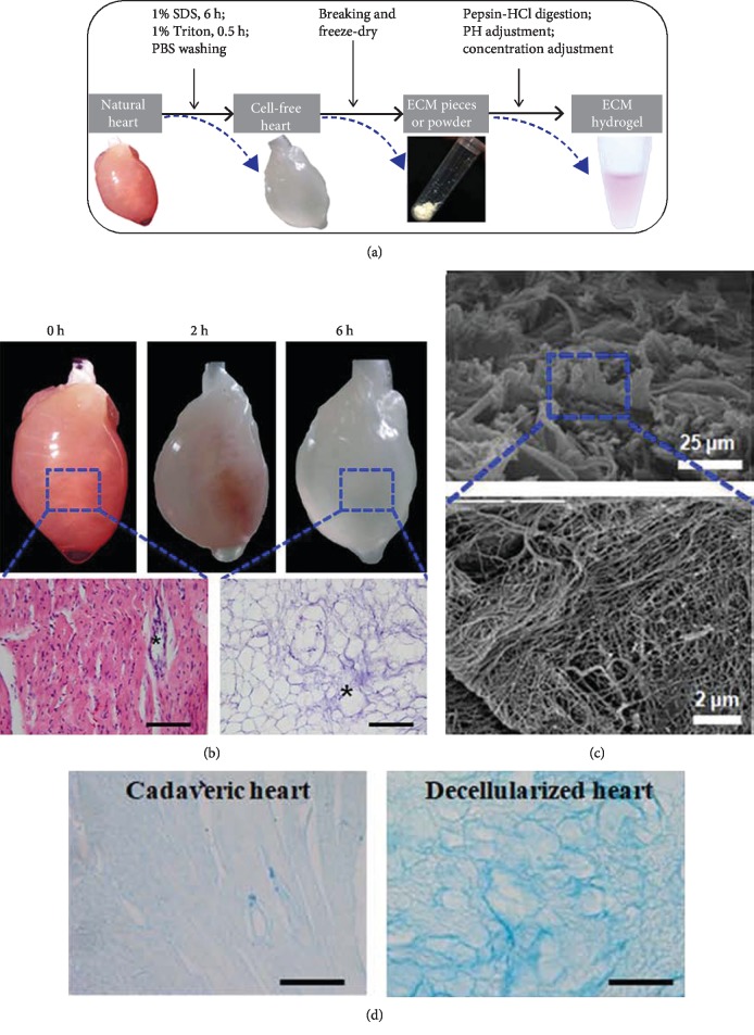
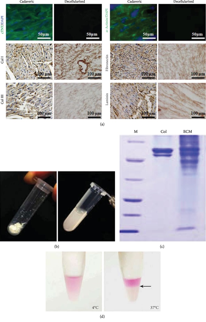
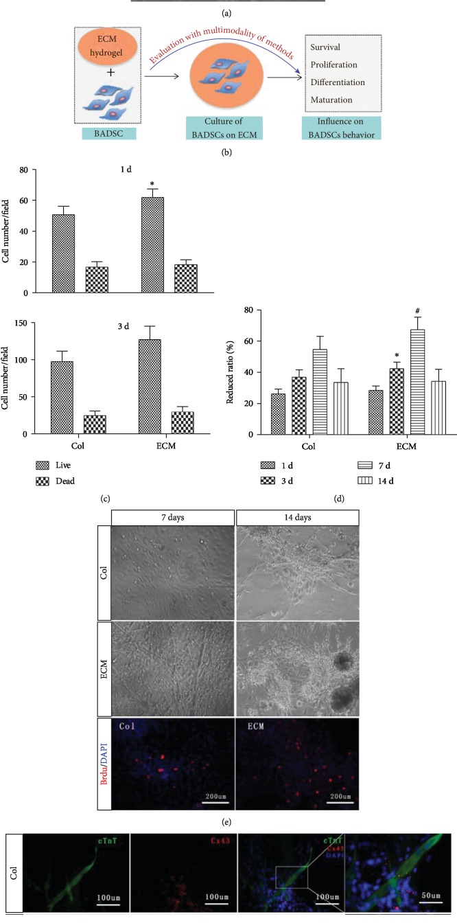
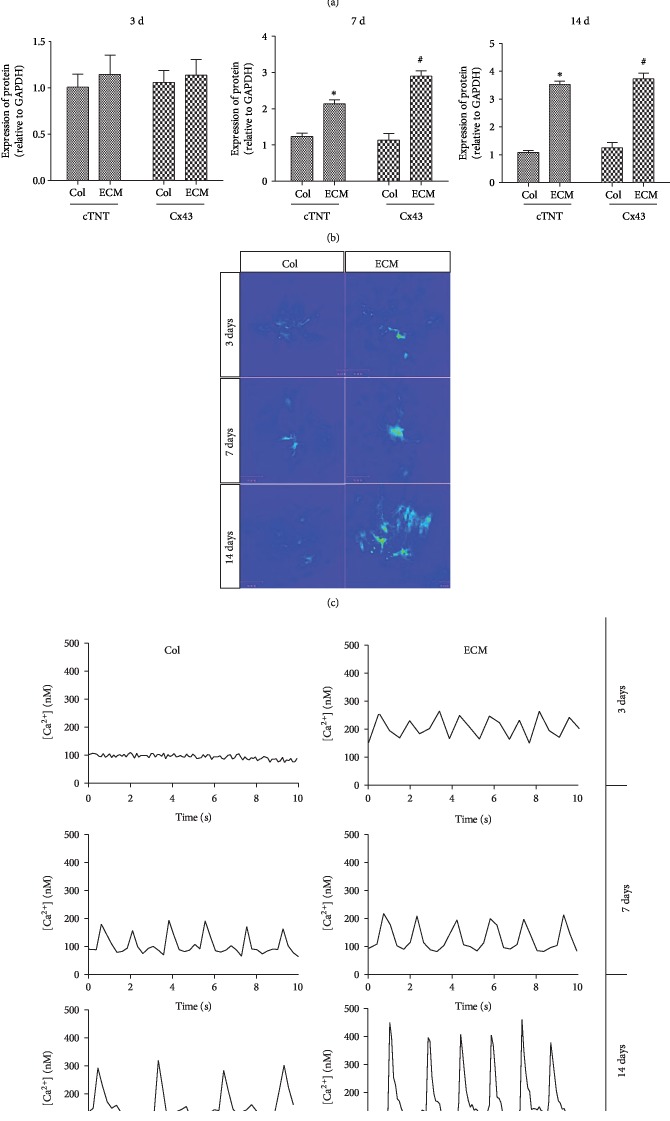
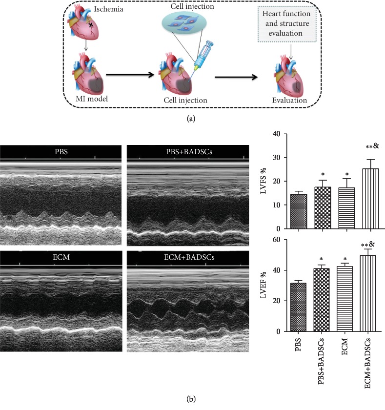
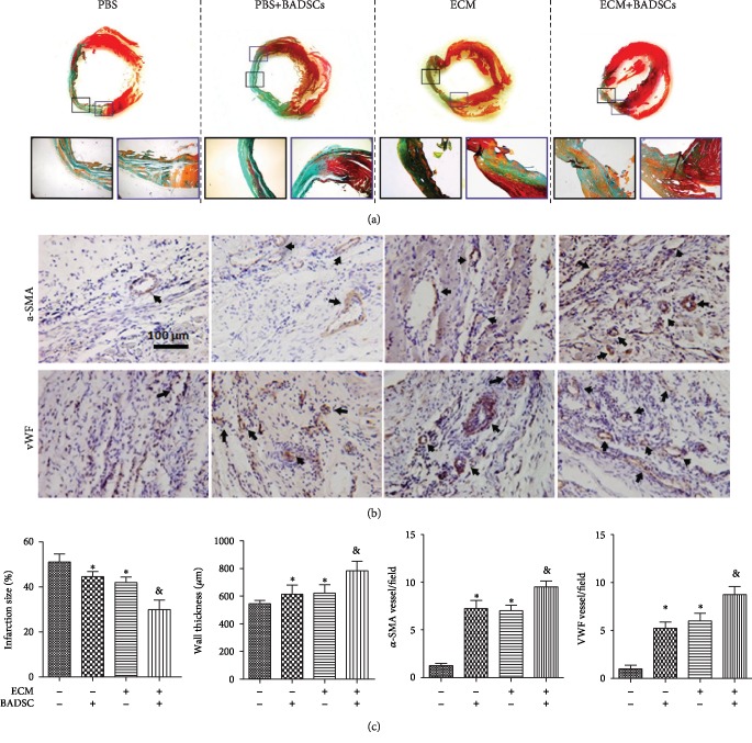
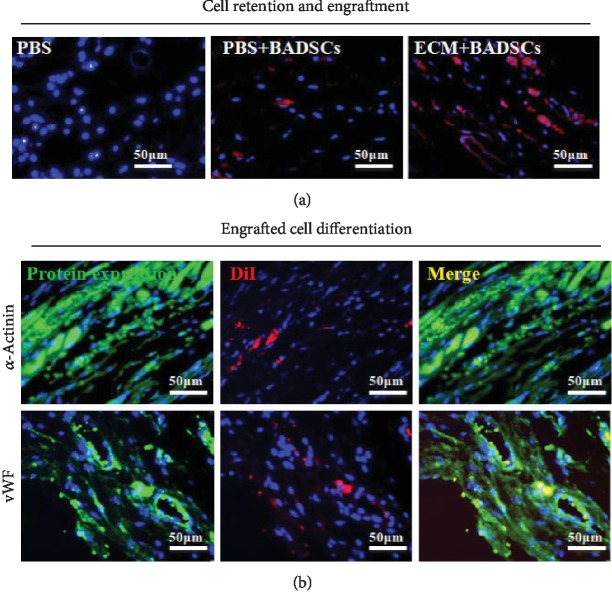
Tissue engineering exploring the combination of scaffolds and seeding cells was proposed as a promising strategy for myocardial repair. However, the therapeutic outcomes varied greatly due to different selection of scaffolds and seeding cells. Herein, the potential of combining bioactive extracellular matrix (ECM) hydrogels and high cardiomyogenic seeding cells was explored for myocardial repair in vitro and in vivo. Temperature-sensitive ECM hydrogels were prepared from decellularized rat hearts, and cardiomyogenic seeding cells were isolated from brown adipose (brown adipose-derived stem cells (BADSCs)). The in vitro studies demonstrated that ECM hydrogel significantly supported the proliferation and cardiomyogenic differentiation of BADSCs. Importantly, the function and maturation of BADSC-derived cardiomyocytes were also promoted as evidenced by Ca2+ transient's measurement and protein marker expression. After myocardial transplantation, the combination of BADSCs and ECM hydrogels significantly preserved cardiac function and chamber geometry compared with BADSCs or ECM hydrogels alone. Meanwhile, the ECM hydrogel also enhanced BADSC engraftment and myocardial regeneration in vivo. These results indicated that heart-derived ECM hydrogels exerted significant influence on the fate of cardiomyogenic cells toward benefiting myocardial repair, which may explain the enhanced stem cell therapy by the scaffold. Collectively, it indicated that the combination of ECM hydrogel and the cardiomyogenic cells may represent a promising strategy for cardiac tissue engineering.


Conflict of interest statement

All authors claim that there are no known conflicts of interest associated with this publication.

Figures

Figure 1
Figure 1
Preparation of decellularized heart ECM. (a) The schematic procedures of preparing decellularized heart hydrogels. (b) Observation of decellularized heart and H&E staining. (c) Scanning electronic observation of decellularized ECM. (d) Alcian Blue staining of natural and decellularized hearts.
Figure 2
Figure 2
Characterization of decellularized heart matrix and preparation of ECM gel. (a) Immunostaining against cardiac markers, cTNT, and α-actinin, as well as extracellular matix components, 8 Col I, fibronectin, Col III, and laminin. (b) Preparation of ECM gel. (c) SDS-PAGE analyzing the components of ECM hydrogel. (d) Thermosensitive feature of the ECM hydrogel.
Figure 3
Figure 3
Evaluation of ECM on the BADSC proliferation and differentiation. (b) The schematic summary of evaluating the influence of ECM hydrogel on BADSC behaviors. (c) Live and dead cell number counting after 1 day and 3 days of culture. (d) Alamar Blue assays demonstrated that both ECM and Col I could promote the proliferation of BADSCs. (e) Morphological observation of BADSCs with time grown on Col I or ECM hydrogel, as well as BrdU labeling of BADSCs at 3 days after seeding. (f) Immunofluorescent staining of cTnT (green) and CX43 (red) expression in BADSCs. p < 0.05 and #p < 0.01 for “ECM vs. Col” at the same time points.
Figure 4
Figure 4
Cardiac differentiation and maturation of BADSCs on ECM-coated substrates. (a) Western blotting detecting the expression of cardiac markers cTnT and CX43. (b) Quantitative analysis of protein expression in BADSCs. (c, d) Ca2+ transient imaging of BADSCs on Col I and ECM-coated substrates. p < 0.05 and #p < 0.01 for “ECM vs. Col” at the same time points.
Figure 5
Figure 5
Cell transplantation and functional evaluation. (a) The schematic summary of MI preparation and cell transplantation. (b) Heart function evaluation by echocardiogram and quantitative analysis of functional parameters, LVFS and LVEF. p < 0.05 compared with control group; p < 0.01 compared with control group; &p < 0.05 compared with the BADSC and ECM alone groups.
Figure 6
Figure 6
Histological examination of injured heart. (a) Masson's trichrome staining. (b) The immunohistochemical staining of vWF and α-SMA for microvessel density analysis in the myocardial infarction sites. Black arrows indicated positively stained vessels. (c) Quantitative analysis for infarct size, wall thickening, and vascular densities in infacted areas. p < 0.05 and ∗∗p < 0.01 for “BADSCs or ECM vs. Col” and &p < 0.01 for “BADSCs or ECM vs. BADSCs+ECM”.
Figure 7
Figure 7
In vivo survival and cardiac differentiation of BADSCs. (a) Histological examination of DiI-labeled BADSCs 4 weeks after implantation (b) and immunofluorescent staining against α-actinin and vWF in the ECM+BADSC group.

References

    1. Go A. S., Mozaffarian D., Roger V. L., et al. Heart disease and stroke statistics—2013 update: a report from the American Heart Association. Circulation. 2013;127:e6–e245. - PMC - PubMed
    1. Mozaffarian D., Benjamin E. J., Go A. S., et al. Heart disease and stroke statistics—2016 update: a report from the American Heart Association. Circulation. 2016;133(4):e38–e360. doi: 10.1161/cir.0000000000000350. - DOI - PubMed
    1. Pillai I. C., Li S., Romay M., et al. Cardiac fibroblasts adopt osteogenic fates and can be targeted to attenuate pathological heart calcification. Cell Stem Cell. 2016;20(2):218–232.e5. doi: 10.1016/j.stem.2016.10.005. - DOI - PMC - PubMed
    1. Braunwald E. The war against heart failure: the Lancet lecture. The Lancet. 2015;385(9970):812–824. doi: 10.1016/s0140-6736(14)61889-4. - DOI - PubMed
    1. Srivastava D., Ivey K. N. Potential of stem-cell-based therapies for heart disease. Nature. 2006;441(7097):1097–1099. doi: 10.1038/nature04961. - DOI - PubMed

LinkOut - more resources