. 2014;1(1):7-14.
doi: 10.15406/jhvrv.2014.01.00003. Epub 2014 May 23.

Noncontiguous Protein Interaction Domains in Osteopontin Contribute to Enhance HIV-1 Replication

Affiliations

Noncontiguous Protein Interaction Domains in Osteopontin Contribute to Enhance HIV-1 Replication

Caitlin Eger et al. J Hum Virol Retrovirol. 2014.

Abstract

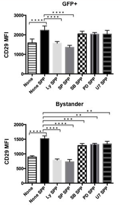
Osteopontin (OPN) is a proinflammatory cytokine produced by T-cells, macrophages, osteoclasts, and several other cell types, which confers immunity to many intracellular pathogens. OPN was first identified as an early marker of cellular activation of T-lymphocytes and subsequently was shown to play a role in cancer through its ability to promote cell survival and inflammation. OPN levels are elevated in the plasma and cerebrospinal fluid of HIV-infected individuals and even more so in those suffering from HIV-related neurocognitive impairment. The infiltration of monocytes and macrophages both infected and uninfected into the brain is the first step in HIV pathogenesis of the central nervous system. Inhibition of OPN in macrophages significantly impairs HIV replication. In an effort to identify and understand the role of OPN in the neuropathogenesis of HIV infection, we are using a combination of in vitro, ex vivo and in vivo approaches. In this study we have used a molecular approach and a surrogate cell culture model to identify the domains of OPN that are required to enhance HIV replication. We found that N- and C-terminal fragments, encoding multiple motifs including sequences involved in binding integrins and CD44, a domain know to promote adhesion contribute to OPN's ability to increase HIV replication. Use of inhibitors against c-Jun N-terminal kinase (JNK) and phosphoinositide 3-kinase (PI-3K) impaired the ability of OPN to increase the integrin subunit 1 or CD29 on the surface of HIV-infected and bystander cells. These results suggest that multiple OPN-regulated cellular pathways are commandeered by HIV to promote productive replication and cell-to-cell spread.

Keywords: Beta-1 integrin; CD29; CD44; JNK; Phosphoinositide-3 kinase; Secreted phosphoprotein-1; Syncytia.


Figures

Figure 1:
Figure 1:
Schematic diagram of SPP1 deletion mutants generated by PCR and cloned into a mammalian expression vector. Osteopontin is encoded by the SPP1 gene and contains several motifs and domains: signal peptide (orange), sites for N--linked glycosylation (pink), an aspartic acid rich domain (green), sequences involved in integrin binding (turquoise and blue), cleavage by thrombin (black) or metalloproteinases (brown), calcium binding (yellow) and heparin and CD44--binding (white). The names of the truncation mutants are indicated beside each construct.
Figure 2a:
Figure 2a:
Reduction in HIV promoter activity with N--terminal truncation of SPP1. The mean fluorescent intensity (MFI) of GFP is an indirect measure of HIV--LTR promoter activity. One--way ANOVA with significance of p<.05 was used. **** p<.0001, *** p<.0005, **p<.005, * p<.05
Figure 2b:
Figure 2b:
SPP1wt and truncation mutants increase CD29 β 1 integrin subunit on HIV--infected compared to bystander GFP negative cells. The mean fluorescent intensity (MFI) of CD29, a measure of the density of the receptor on the cell surface is shown for cells not expressing SPP1 (None) and for each of the truncation mutants. One--way ANOVA with significance of p<.05 was used. **** p<.0001, *** p<.0005, **p<.005, * p<.05
Figure 3:
Figure 3:
SPP1 deletions enhance cell--to--cell fusion. Images of TZM--bl cells transfected with SPP1wt (OPN) or the indicated SPP1 deletion mutants and infected with HIVSF162--R3 GFP after 2--3 days post--infection are shown.
Figure 4:
Figure 4:
Inhibition of PI3K or JNK signaling inhibits the ability of SPP1 to increase CD29 β1 integrin subunit expression on HIV--infected (GFP+) and bystander cells. TZM--bl cells were first transfected with the SPP1wt expression plasmid followed by overnight infection with HIVSF162--R3 GFP. The next day the medium was changed and the inhibitors U73122 (1μM, U7), PD98059 (10μM, PD), SB203580 (10μM, SB), SP600125 (10μM, SP) and LY294002 (50μM, Ly) were added. The cells were harvested 3 days later for flow cytometric analyses of CD29 expression. One--way ANOVA with significance of p<.05 was used. **** p<.0001, *** p<.0005, **p<.005, * p<.05.

References

    1. Brown A, Islam T, Adams R, Nerle S, Kamara M, et al. (2011) Osteopontin enhances HIV replication and is increased in the brain and cerebrospinal fluid of HIV-infected individuals. J Neurovirol 17(4): 382–392. - PMC - PubMed
    1. Roberts ES, Zandonatti MA, Watry DD, Madden LJ, Henriksen SJ, et al. (2003) Induction of pathogenic sets of genes in macrophages and neurons in NeuroAIDS. Am J Pathol 162(6): 2041–2057. - PMC - PubMed
    1. Burdo TH, Ellis RJ, Fox HS (2008) Osteopontin is increased in HIV-associated dementia. J Infect Dis 198(5): 715–722. - PMC - PubMed
    1. Burdo TH, Wood MR, Fox HS (2007) Osteopontin prevents monocyte recirculation and apoptosis. J Leukoc Biol 81(6): 1504–1511. - PMC - PubMed
    1. Wang KX, Denhardt DT (2008) Osteopontin: Role in immune regulation and stress responses. Cytokine Growth Factor Rev 19(5–6): 333–345. - PubMed

LinkOut - more resources