Review
. 2020 Jun;47(6):1538-1551.
doi: 10.1007/s00259-019-04603-1. Epub 2019 Nov 27.

Atherosclerosis imaging with 18F-sodium fluoride PET: state-of-the-art review

Affiliations
Review

Atherosclerosis imaging with 18F-sodium fluoride PET: state-of-the-art review

Poul F Høilund-Carlsen et al. Eur J Nucl Med Mol Imaging. 2020 Jun.

Abstract

Purpose: We examined the literature to elucidate the role of 18F-sodium fluoride (NaF)-PET in atherosclerosis.

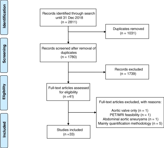
Methods: Following a systematic search of PubMed/MEDLINE, Embase, and Cochrane Library included articles underwent subjective quality assessment with categories low, medium, and high. Of 2811 records, 1780 remained after removal of duplicates. Screening by title and abstract left 41 potentially eligible full-text articles, of which 8 (about the aortic valve (n = 1), PET/MRI feasibility (n = 1), aortic aneurysms (n = 1), or quantification methodology (n = 5)) were dismissed, leaving 33 published 2010-2012 (n = 6), 2013-2015 (n = 11), and 2016-2018 (n = 16) for analysis.

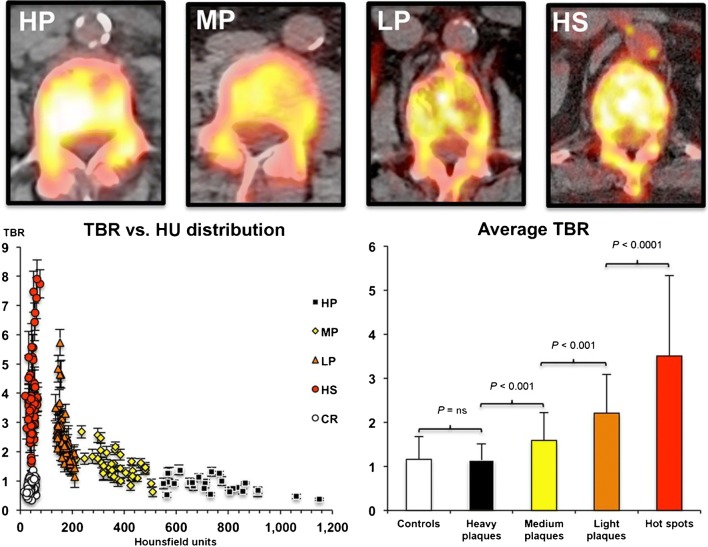
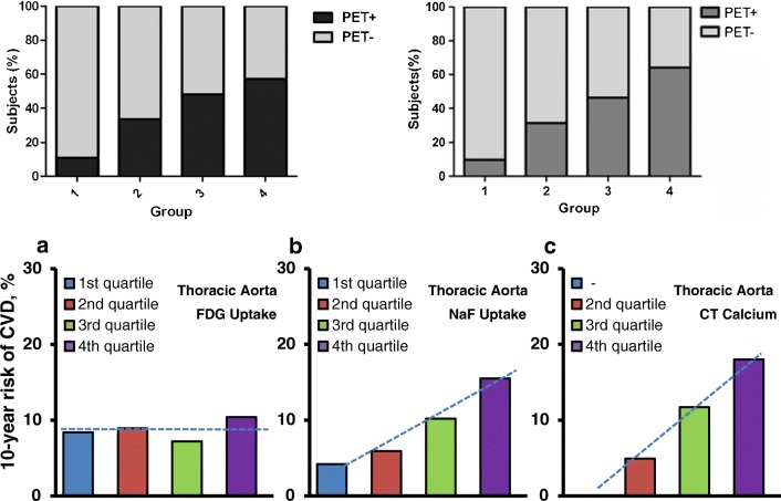
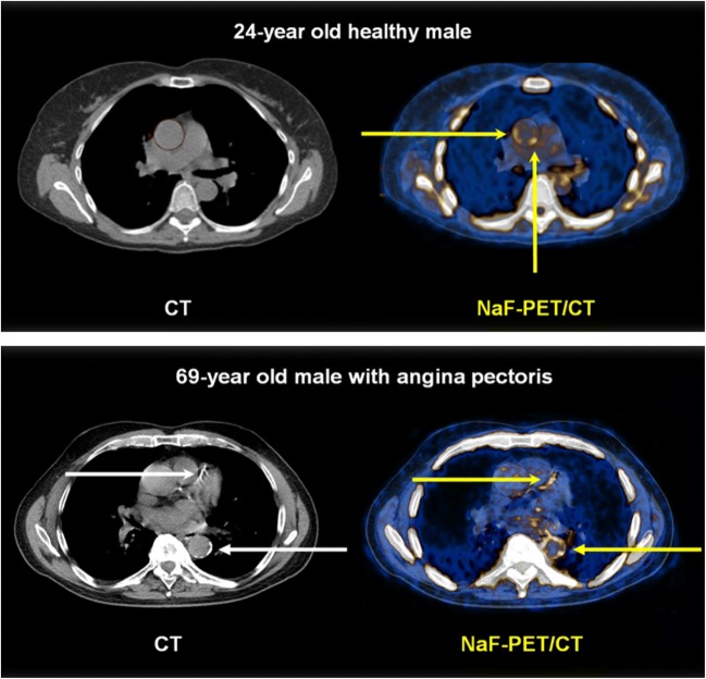
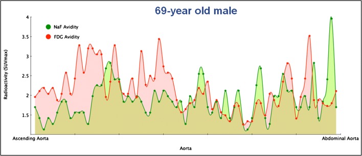
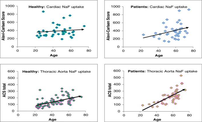
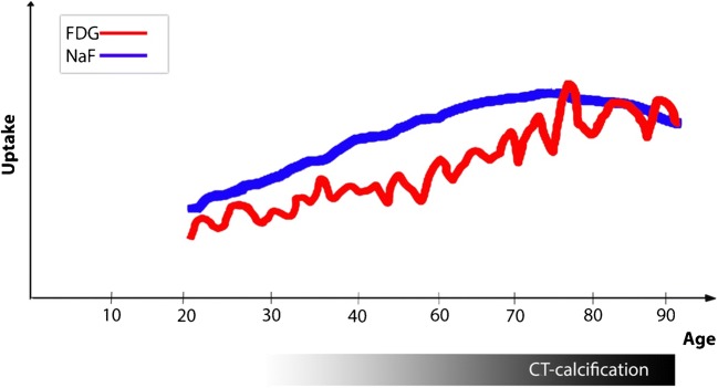
Results: They focused on coronary (n = 8), carotid (n = 7), and femoral arteries (n = 1), thoracic aorta (n = 1), and infrarenal aorta (n = 1). The remaining 15 studies examined more than one arterial segment. The literature was heterogeneous: few studies were designed to investigate atherosclerosis, 13 were retrospective, 9 applied both FDG and NaF as tracers, 24 NaF only. Subjective quality was low in one, medium in 13, and high in 19 studies. The literature indicates that NaF is a very specific tracer that mimics active arterial wall microcalcification, which is positively associated with cardiovascular risk. Arterial NaF uptake often presents before CT-calcification, tends to decrease with increasing density of CT-calcification, and appears, rather than FDG-avid foci, to progress to CT-calcification. It is mainly surface localized, increases with age with a wide scatter but without an obvious sex difference. NaF-avid microcalcification can occur in fatty streaks, but the degree of progression to CT-calcification is unknown. It remains unknown whether medical therapy influences microcalcification. The literature held no therapeutic or randomized controlled trials.

Conclusion: The literature was heterogeneous and with few clear cut messages. NaF-PET is a new approach to detect and quantify microcalcification in early-stage atherosclerosis. NaF uptake correlates with cardiovascular risk factors and appears to be a good measure of the body's atherosclerotic burden, potentially suited also for assessment of anti-atherosclerotic therapy.

Keywords: 18F-sodium fluoride; Atherosclerosis; Calcification; NaF; PET; Quantification.


Conflict of interest statement

The authors declare that they have no conflict of interest.

Figures

Fig. 1
Fig. 1
PRISMA flow diagram
Fig. 2
Fig. 2
Inverse relation between arterial wall uptake of NaF and CT-visible calcification in the infrarenal aorta. High NaF uptake in aortic lesion without CT-calcification (Hot spots = HS) and decreasing NaF uptake with increasing density of CT-calcifications until same low NaF uptake in high density plaques (Heavy plaques = HP) than in controls (reproduced by permission from reference 24)
Fig. 3
Fig. 3
Arterial mineral deposition increases with age, while regional bone metabolism decreases. Coronal fused 18F-sodium fluoride PET/CT (a) and non-fused PET of the thighs (b) of a 29-year-old woman with low arterial mineral deposition (TBR 1.44) and high bone metabolism (SUVmean 8.6). Coronal fused 18F-sodium fluoride PET/CT (c) and non-fused PET of the thighs (d) of a 83-year-old woman showing high arterial mineral deposition (TBR 2.18) and low bone metabolism (SUVmean 6.7) (reproduced by permission from reference 25)
Fig. 4
Fig. 4
Positive relationship between arterial NaF uptake and cardiovascular risk. Upper panel, left: Relationship between cardiovascular risk factors and presence of NaF uptake in carotid arteries. (reproduced with permission from reference 5). Upper panel, right: Relationship between cardiovascular risk factors and presence of NaF uptake in carotid arteries (reproduced with permission from reference 35). Lower panel: 10-year Framingham risk score in relation to quartiles of (a) thoracic aorta FDG activity, (b) thoracic aorta NaF activity, and (c) thoracic aorta CT calcium burden. The risk is similar in all quartiles of thoracic aorta FDG uptake, but increases linearly with each increasing quartile of thoracic aorta NaF uptake (P < 0.001 for a linear trend) and with each increasing quartile of thoracic aorta CT calcium burden (P < 0.001 for a linear trend) (modified from reference 23)
Fig. 5
Fig. 5
Abnormal arterial wall NaF uptake has been observed down to the age of 20 and is most often present in the absence of CT-calcification. Upper panel: NaF uptake (right) in the thoracic aorta (yellow arrows) of a 24-year-old symptom-free male with no CT-visible calcification (left). Lower panel: 69-year-old male with angina pectoris and CT-visible calcification in the left anterior descending coronary artery and the descending thoracic aorta (white arrows, left) and NaF uptake in the same arteries, incongruent with CT-calcifications and with far greater circumferential extension in the aorta (yellow arrows, right) (images from material of references and 30)
Fig. 6
Fig. 6
NaF uptake is often not confined to identifiable lesions, but varies through each arterial segment. Variation in FDG (red areas) and NaF (green areas) uptake through the entire aorta. The red and green dots are blood background subtracted SUVmax values from horizontal slices of 3.75 mm thickness. Note the outspoken disconcordance and the generally higher FDG uptake in the thoracic part (to the left) and generally higher NaF uptake in the abdominal part of the aorta (to the right) (from the material of reference 23)
Fig. 7
Fig. 7
NaF uptake increases with age, the more so in patients, but with a wide scatter. NaF uptake in the heart and thoracic aorta of healthy control subjects and angina pectoris patients as a function of age. Note the steeper slope in angina pectoris patients. All correlations were statistically significant, but with a large scatter indicating that among both healthy individuals and cardiac patients, there are some individuals with very low and some with very high NaF uptake (from the CAMONA material of references and 23)
Fig. 8
Fig. 8
Hypothetical illustration of the possible time- and age-related relationship among arterial wall FDG uptake, NaF uptake, and CT-visible calcification. The courses in childhood and very high ages are unknown. FDG uptake may be a frequently occurring repetitive process throughout life in response to minor or major arterial injuries [44]. By targeting microcalcification [17], NaF appears to be a more persistent marker of early phase atherosclerosis [4, 15, 24, 30, 36] and, through surface adsorption to macrocalcification [17], to a lesser degree also of still ongoing calcification in CT-visible calcifications [24]. The three processes follow different patterns, a slow and protracted increase of FDG uptake, a similarly early in life occurring NaF uptake that tends to persist and increase for some age decades until it decreases when macrocalcification grows and stabilizes, and finally, and in contrast, CT-detectable calcification that appears later in life and continues with aging (illustration by Dr. Reza Piri, Dept. of Nuclear Medicine, Odense University Hospital, Odense, Denmark)

References

    1. Worlds Health Organization. Cardiovascular disease. WHO 2019. https://www.who.int/cardiovascular_diseases/en/. Accessed 15 Sep 2019.
    1. Yun M, Yeh D, Araujo LI, Jang S, Newberg A, Alavi A. F-18 FDG uptake in the large arteries: a new observation. Clin Nucl Med. 2001;26:314–319. doi: 10.1097/00003072-200104000-00007. - DOI - PubMed
    1. Yun M, Jang S, Cucchiara A, Newberg AB, Alavi A. 18F FDG uptake in the large arteries: a correlation study with the atherogenic risk factors. Semin Nucl Med. 2002;32:70–76. doi: 10.1053/snuc.2002.29279. - DOI - PubMed
    1. Derlin T, Richter U, Bannas P, et al. Feasibility of 18F-sodium fluoride PET/CT for imaging of atherosclerotic plaque. J Nucl Med. 2010;51:862–865. doi: 10.2967/jnumed.110.076471. - DOI - PubMed
    1. Derlin T, Wisotzki C, Richter U, et al. In vivo imaging of mineral deposition in carotid plaque using 18F-sodium fluoride PET/CT: correlation with atherogenic risk factors. J Nucl Med. 2011;52:362–368. doi: 10.2967/jnumed.110.081208. - DOI - PubMed