. 2019 Nov 25;8(12):1508.
doi: 10.3390/cells8121508.

MS CD49d+CD154+ Lymphocytes Reprogram Oligodendrocytes into Immune Reactive Cells Affecting CNS Regeneration

Affiliations

MS CD49d+CD154+ Lymphocytes Reprogram Oligodendrocytes into Immune Reactive Cells Affecting CNS Regeneration

Paweł Piatek et al. Cells. .

Abstract

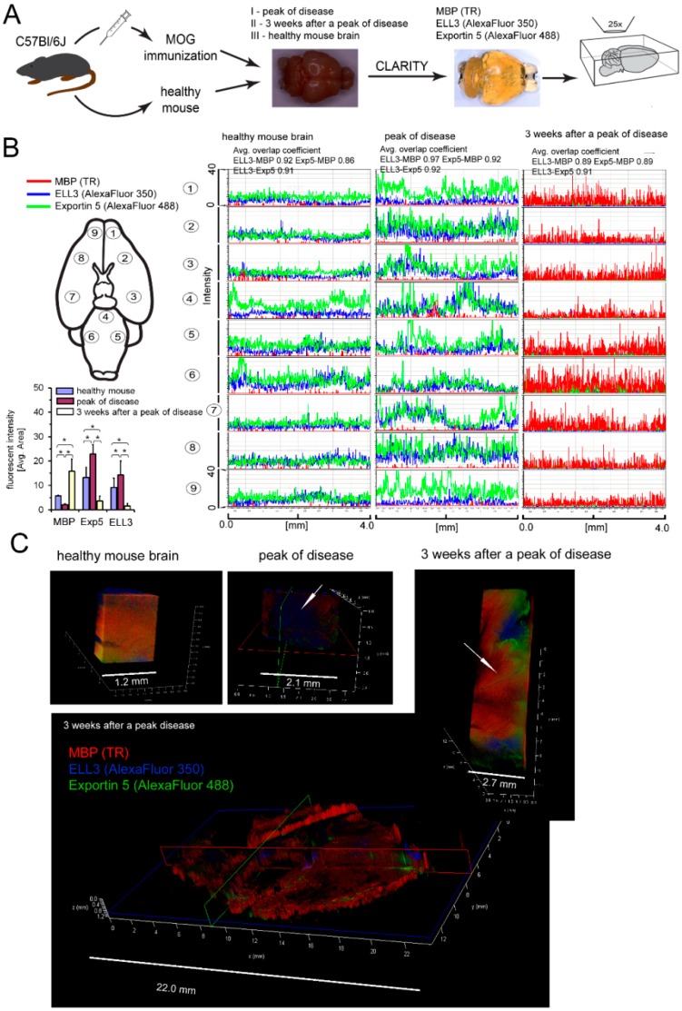
The critical aspect in multiple sclerosis (MS) progression involves insufficient regeneration of CNS resulting from deficient myelin synthesis by newly generated oligodendrocytes (OLs). Although many studies have focused on the role of autoreactive lymphocytes in the inflammatory-induced axonal loss, the problem of insufficient remyelination and disease progression is still unsolved. To determine the effect of myelin-specific lymphocytes on OL function in MS patients and in a mouse model of MS, we cultured myelin induced MS CD49d+CD154+ circulating lymphocytes as well as Experimental Autoimmune Encephalomyelitis (EAE) mouse brain-derived T and memory B cells with maturing oligodendrocyte precursor cells (OPCs). We found that myelin-specific CD49d+CD154+ lymphocytes affected OPC maturation toward formation of immune reactive OLs. Newly generated OLs were characterized by imbalanced myelin basic protein (MBP) and proteolipid protein (PLP) production as well as proinflammatory chemokine/cytokine synthesis. The analysis of cellular pathways responsible for OL reprogramming revealed that CD49d+CD154+ lymphocytes affected miRNA synthesis by dysregulation of polymerase II activity. miR-665 and ELL3 turned out to be the main targets of MS myelin-specific lymphocytes. Neutralization of high intracellular miR-665 concentration restored miRNA and MBP/PLP synthesis. Together, these data point to new targets for therapeutic intervention promoting CNS remyelination.

Keywords: multiple sclerosis; myelin-specific lymphocytes; oligodendrocyte precursor cells; remyelination.


Conflict of interest statement

The authors declare no conflict of interest.

Figures

Figure 1
Figure 1
Relapsing-remitting multiple sclerosis (RR-MS) CD49d+CD154+ lymphocytes affect maturing oligodendrocyte precursor cells (OPCs), resulting in dysregulation of myelin production by mature oligodendrocytes (OLs). (A) RR-MS CD49d+CD154+, contrary to healthy control (HC) lymphocytes, affected myelin basic protein (MBP) and proteolipid protein (PLP) but not myelin oligodendrocyte glycoprotein (MOG) synthesis by human OPCs (hOPCs). (B) MBP/PLP index more clearly exhibited disproportion in myelin protein synthesis. (C) Immunocytochemical (ICC) analysis of nestin, O4 and GFAP expression in hOPCs demonstrated that RR-MS CD49d+CD154+ lymphocytes did not affect hOPC maturation, but caused dysregulation of MBP and PLP synthesis by mature OLs. All data are presented as means ± SD.
Figure 2
Figure 2
Mouse brain-infiltrating mononuclear cells (BMCs) affect myelin protein synthesis by mouse OPCs (mOPCs). (A) BMCs from Experimental Autoimmune Encephalomyelitis (EAE) mice in the third week after the peak of the disease (remyelination phase) were cocultured with OPCs isolated from the healthy mouse newborns (mOPCs). (A1, left panel) Phenotypic analysis of BMCs (region R1) divided into B cells (B220, region R2) and T cells (TCR-β, region R3). (A1, right panel) CD3+ and CD19+ cell rate analysis in isolated BMCs. (A2) Memory B cells (mCD19+) were positively selected, and unlabeled cells additionally sorted by negative selection into CD3+ lymphocytes from BMCs. (B, upper panel) In contrast to immature cells, mature OLs exhibited MBP/PLP/MOG expression (markers of the late stage of differentiation), O4 expression (marker of mature cells), and negative signal for nestin (marker of OL precursor cells) and GFAP (astrocyte-specific marker). (B, low panel) EAE CD3+/mCD19+ and CD3+ cells alone down- PLP and upregulated MBP expression, but did not affect O4 or nestin. Maturation process was associated with formation of actin-like microfilaments (labelled with phalloidin). Confocal z-stack analysis confirmed that EAE CD3+/mCD19+ BMC-derived cells did not affect O4 accumulation in the intracellular space and microfilament formation during OPC maturation. (C) mRNA myelin protein analysis proved that EAE CD3+/mCD19+ and CD3+ BMC-derived cells up- MBP and downregulated PLP, but had no influence on MOG synthesis. All data are presented as means ± SD.
Figure 3
Figure 3
EAE BMC-derived cells as well as RR-MS CD49d+CD154+ lymphocytes affect miRNA synthesis, but have no direct effect on the transcriptional factors. (A) EAE BMC-derived cells did not change mRNA expression of transcription factors responsible for adult OPC activation (green font) as well as differentiation and myelin sheaths formation (red font). (B) RR-MS CD49d+CD154+ lymphocytes as well as CD3+/mCD19+ and CD3+ EAE BMC-derived cells affected intracellular miRNA concentrations during OPC maturation. Data are presented as means ± SD. (C) Bioinformatic analysis of genes responsible for mRNA binding and miRNA processing point to the polymerase II elongation factor (ELL3). From the total of 33,000 genes, those mostly affected during physiological OPC maturation and associated with mRNA binding or engagement in miRNA synthesis were selected. 43 genes from this group, with statistically significant fluctuations in the presence of CD3+/mCD19+ EAE BMC-derived cells were selected. Twenty-three were upregulated (red font) and 20 downregulated (blue font). The most significant alterations were observed in ELL3 expression. (D) The validation of ELL3 and the other miRNA processing proteins by qPCR points to Exp5, DICER1, and Ago2, which were reduced in maturating OPCs in the presence of CD3+/mCD19+ EAE BMC-derived cells.
Figure 4
Figure 4
Pathological SM lesions in the period between the active and remission phase of disease characterized by changeable ELL3 and Exp5 expression which correspond with MBP synthesis. (A) Colocalization of ELL3 and Exp5 miRNA processing proteins in the region of the demyelinating and remyelinating plaques during the course of disease using the whole imaging of mouse EAE brain by CLARITY. (B, left upper panel) The fluorescent intensity of MBP (red pseudocolor), ELL3 (blue) and Exp5 (green) was measured in nine regions of each investigated brains in every 4.0 mm of tissue thickness. (B, left lower panel) Downregulation of ELL3 synthesis is associated with reduced Exp5 expression. Data are presented as means ± SD. (B, right panel) High colocalization of florescent signals (average overlap coefficients factor > 0.91) between MBP-ELL3, MBP-Exp5 and ELL3-Exp5 in all analyzed regions suggested that intensity of miRNA synthesis and its transport to cytoplasm was correlated with MBP expression. (C, left and middle pictures). In contrast to the healthy mouse, in the peak of EAE, demyelination regions were characterized by the loss of MBP and high ELL3 and Exp5 expressions (an arrow points demyelinating plaque). (C, lower and right pictures) After three weeks, this process was reversed as MBP was highly overexpressed and ELL3 and Exp5 were downregulated (an arrow points remyelinating region).
Figure 5
Figure 5
RR-MS myelin-specific CD49d+CD154+ lymphocytes interfere with miRNA profiling in maturing OPCs. (A). Heat map revealed a specific miRNA profile for every cell culture experimental settings. Based on the differences between the effect of RR-MS and HC CD49d+CD154+ lymphocytes on OPCs, three clusters were selected (red, green, and blue frames). The most significant differences were observed in the cluster with hsa-miR-21-5p, hsa-miR-221-5p, hsa-miR-222-3p, hsa-miR-21-3p hsa-miR-665 (red frame), and hsa-miR-212-3p (blue frame). The data was normalized with the trimmed mean of M-values method and converted to a log2 scale. Red represents an expression level above the mean, while green one level below. (B) Validation of selected miRNA by copy transcript estimation using ddPCR analysis in additional cohort patients. Data are presented as means ± SD.
Figure 6
Figure 6
The crucial role of hsa-miR-665 and hsa-miR-21-3p in myelin protein dysregulations in hOPCs exposed to RR-MS CD49d+CD154+ lymphocytes. (A,B) Neutralization of hsa-miR-665 but not hsa-miR-21-3p, improved ELL3, PLP mRNA synthesis, and reduced MBP mRNA expression. (C) Neutralization of hsa-miR-665 but not hsa-miR-21-3p completely reversed the effect of RR-MS CD49d+CD154+ lymphocytes on miRNA synthesis by hOPCs. All data are presented as means ± SD.

Similar articles

Cited by

References

    1. Lublin F.D., Reingold S.C., Cohen J.A., Cutter G.R., Sørensen P.S., Thompson A.J., Wolinsky J.S., Balcer L.J., Banwell B., Barkhof F., et al. Defining the clinical course of multiple sclerosis: The 2013 revisions. Neurology. 2014;83:278–286. doi: 10.1212/WNL.0000000000000560. - DOI - PMC - PubMed
    1. Bramow S., Frischer J.M., Lassmann H., Koch-Henriksen N., Lucchinetti C.F., Sørensen P.S., Laursen H. Demyelination versus remyelination in progressive multiple sclerosis. Brain. 2010;133:2983–2998. doi: 10.1093/brain/awq250. - DOI - PubMed
    1. Chang A., Tourtellotte W.W., Rudick R., Trapp B.D. Premyelinating oligodendrocytes in chronic lesions of multiple sclerosis. N. Engl. J. Med. 2002;346:165–173. doi: 10.1056/NEJMoa010994. - DOI - PubMed
    1. Barnett M.H., Prineas J.W. Relapsing and remitting multiple sclerosis: Pathology of the newly forming lesion. Ann. Neurol. 2004;55:458–468. doi: 10.1002/ana.20016. - DOI - PubMed
    1. Ffrench-Constant C., Raff M.C. Proliferating bipotential glial progenitor cells in adult rat optic nerve. Nature. 1986;319:499–502. doi: 10.1038/319499a0. - DOI - PubMed

Publication types