. 2020 Jan-Dec;12(1):1684749.
doi: 10.1080/19420862.2019.1684749.

Development of a high yielding expression platform for the introduction of non-natural amino acids in protein sequences

Affiliations

Development of a high yielding expression platform for the introduction of non-natural amino acids in protein sequences

Gargi Roy et al. MAbs. 2020 Jan-Dec.

Abstract

The ability to genetically encode non-natural amino acids (nnAAs) into proteins offers an expanded tool set for protein engineering. nnAAs containing unique functional moieties have enabled the study of post-translational modifications, protein interactions, and protein folding. In addition, nnAAs have been developed that enable a variety of biorthogonal conjugation chemistries that allow precise and efficient protein conjugations. These are being studied to create the next generation of antibody-drug conjugates with improved efficacy, potency, and stability for the treatment of cancer. However, the efficiency of nnAA incorporation, and the productive yields of cell-based expression systems, have limited the utility and widespread use of this technology. We developed a process to isolate stable cell lines expressing a pyrrolysyl-tRNA synthetase/tRNApyl pair capable of efficient nnAA incorporation. Two different platform cell lines generated by these methods were used to produce IgG-expressing cell lines with normalized antibody titers of 3 g/L using continuous perfusion. We show that the antibodies produced by these platform cells contain the nnAA functionality that enables facile conjugations. Characterization of these highly active and robust platform hosts identified key parameters that affect nnAA incorporation efficiency. These highly efficient host platforms may help overcome the expression challenges that have impeded the developability of this technology for manufacturing proteins with nnAAs and represents an important step in expanding its utility.

Keywords: Antibody drug conjugate; IgG; PylRS; non-natural amino acid; tRNA.


Figures

Figure 1.
Figure 1.
mRNA reporter assay for screening amber suppression positive stable isolate hosts. (a) Schematic representation of the process used to generate and identify amber suppression competent cells. CHO cells were first transfected with a plasmid encoding PylRS and tRNApyl and subjected to a selection step by growth in puromycin-containing medium. Surviving isolates were transfected with mRNA encoding RFPambGFP and cells were exposed to AzK. Cells capable of efficient amber codon suppression (positive) express an RFP-GFP fusion and are positive for both fluorophores. Cells lacking amber suppression activity (negative) express only RFP. (b) A stable isolate identified as positive for amber suppression activity was transfected with the RFPambGFP mRNA and incubated in the presence or absence of AzK. Cells were imaged by confocal microscopy and flow cytometry. Amber suppression and incorporation of AzK was observed in presence of PylRS and tRNApyl. No GFP expression was observed in absence of AzK.
Figure 2.
Figure 2.
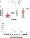
Screening criteria for selection of amber suppression efficient hosts. (a) Structure of AzK. (b) Ranking of platform hosts based on amber suppression activity determined by the ratio of mean fluorescence intensity of GFP to RFP (GFP/RFP) or (c) based on the percentage of the population showing dual fluorescence protein expression. The relative ranking of the three top hosts based on IgG expression are indicated with pink (27), blue (38) and green markers (43). (d) Isolates identified as positive for amber suppression (n = 50) were assessed for amber codon dependent IgG expression. Candidate hosts were transfected with IgGamb and four pools from each candidate assessed for IgGamb titer in the presence of AzK. Titer values for each pool were plotted with median expression shown. Host candidates 27, 38 and 43 showed the highest IgGamb expression levels and were selected for further evaluation.
Figure 3.
Figure 3.
Correlation between PylRS and tRNApyl copy numbers and IgG Titer. (a) Correlation between PylRS gene copy number and IgG titer of the isolated host platforms. The red marker indicates host 43. (b) Correlation between tRNApyl copy number and IgG titer of the isolated host platforms. The red marker indicates host 43. (c) Correlation between PylRS gene copy number and IgG titer of 10 clonal hosts derived from host 43 (solid red). (d) Correlation between tRNApyl copy number and IgG titer of the 10 clonal hosts derived from host 43 (solid red).
Figure 4.
Figure 4.
Cell growth and productivity profiles of different IgG-expressing stable lines containing different levels of amber suppression activity. Expression cell lines derived from C13–27, and −43 were generated and subjected to a 14-day fed-batch expression. For each cell line we determined: (a) viable cell densities, (b) cell viability, and (c) full length and truncated IgG titer. (d) Crude supernatants from each fed-batch culture were resolved under non-reducing condition by SDS-PAGE and analyzed by Western blotting using an antibody that recognizes the truncated and full-length IgG. The positions of full-length and truncated IgG heavy chain, light chain (LC) and light chain dimers (2xLC) are indicated. The efficiency of amber suppression in each culture was calculated after quantifying full-length and truncated IgG levels by surface layer interferometry.
Figure 5.
Figure 5.
Screening strategy yields efficient PylRSY306A mutant host. (a) Distribution of IgG titer in stable pools generated in Y306A and tRNApyl expressing platform hosts. Best performing hosts are shown with colored markers. (b) Median IgG titers of stable pools generated from WT and Y306A host platforms were plotted. Y306A host cells show improved median expression levels of full-length IgGamb.
Figure 6.
Figure 6.
Monitoring of cell performance, amber suppression efficiency and titer distribution during perfusion bioreactor process development. Viable cell density (a), cell viability (b), full-length IgG titer (c) and amber suppression efficiency (d) were measured in anti-HER2 IgG-expressing cell lines S/AzK, V/AzK, V/CpHK and V/SCpHK in perfusion mode. S/AzK is expressed in WT PylRS/tRNApyl platform host while V/AzK, V/CpHK and V/SCpHK represents a cell line derived from the Y306A/tRNApyl platform host and induced with AzK, CpHK and SCpHK nnAAs.
Figure 7.
Figure 7.
Functional assessment of nnAA bearing antibodies. Antibodies purified from each of the perfusion cultures were purified and characterized for conjugation efficacy and ADC function. (A) Affinity-purified antibodies were conjugated to tubulysin warheads containing alkyne- (DBCO; for AzK IgGs) or maleimide- (for SCpHK and CpHK) bearing linkers. Unconjugated (-) and conjugated samples were resolved by SDS-PAGE and stained with Coomassie. A retardation in gel mobility is observed in the HC, but not the LC, of conjugated samples, suggesting near-complete conjugate formation. (b) Reduced antibodies and ADCs were analyzed by mass spectrometry. ADCs show an intact HC mass increase corresponding to the mass of the DBCO-tubulysin (1074.6 Da) or maleimide-tubulysin (1091.61 Da) adducts indicating the addition of a single payload to the HC. No unreacted material was observed in ADC samples. C. and D. The constructed ADCs were functionally tested in an in vitro cytotoxicity assay. Antibodies and ADCs were incubated with HER2 positive (Sk-Br-3) (c) and negative (MDA-MB-468) (d) cell lines and their viabilities were assessed. V/CpHK (-) in C represents HER2 mAb without a toxic payload. All ADCs show potent and specific on target killing.
Figure 8.
Figure 8.
Intracellular localization of PylRS. (a) Localization of recombinant PylRS by confocal microscopy in primary isolates of IgG expressors in host 43 platform hosts. Cells were fixed and stained with anti-FLAG antibodies (PylRS) and the membrane protein Atp1p. DAPI was used to stain the nuclei. PylRS seems to localize to the cytoplasm of cells. (b) To examine whether PylRS is secreted, 50 µg of whole-cell lysates (L), 1 µg crude supernatants (S), and 1 ug of purified IgG derived from host 43 expressers were resolved by SDS-PAGE and examined by Western blotting. FLAG-tagged PylRS (upper panel) was detected using anti-FLAG antibody. IgG HC and LC were examined as loading controls (lower panel).

References

    1. Dall’Acqua WF, Kiener PA, Wu H.. Properties of human IgG1s engineered for enhanced binding to the neonatal Fc receptor (FcRn). J Biol Chem. 2006;281:23514–17. doi:10.1074/jbc.M604292200. - DOI - PubMed
    1. Thakur A, Huang M, Lum LG.. Bispecific antibody based therapeutics: strengths and challenges. Blood Rev. 2018;32:339–47. doi:10.1016/j.blre.2018.02.004. - DOI - PubMed
    1. Sliwkowski MX, Mellman I. Antibody therapeutics in cancer. Science. 2013;341:1192–98. doi:10.1126/science.1241145. - DOI - PubMed
    1. Zhu GD, Fu YX. Design of next generation antibody drug conjugates. Yao Xue Xue Bao. Acta Pharmaceutica Sinica. 2013;48:1053–70. - PubMed
    1. Hamblett KJ, Senter PD, Chace DF, Sun MM, Lenox J, Cerveny CG, Kissler KM, Bernhardt SX, Kopcha AK, Zabinski RF, et al. Effects of drug loading on the antitumor activity of a monoclonal antibody drug conjugate. Clin Can Res. 2004;10:7063–70. doi:10.1158/1078-0432.CCR-04-0789. - DOI - PubMed

MeSH terms

LinkOut - more resources