. 2019 Nov 27;15(1):421.
doi: 10.1186/s12917-019-2171-7.

Development and comparison of enzyme-linked immunosorbent assays based on recombinant trimeric full-length and truncated spike proteins for detecting antibodies against porcine epidemic diarrhea virus

Affiliations

Development and comparison of enzyme-linked immunosorbent assays based on recombinant trimeric full-length and truncated spike proteins for detecting antibodies against porcine epidemic diarrhea virus

Chia-Yu Chang et al. BMC Vet Res. .

Abstract

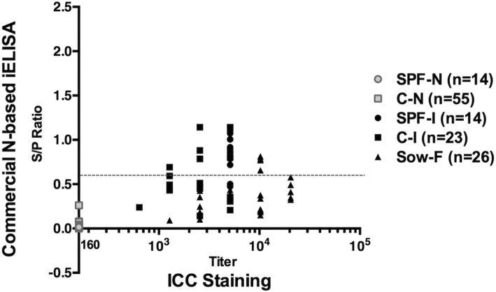
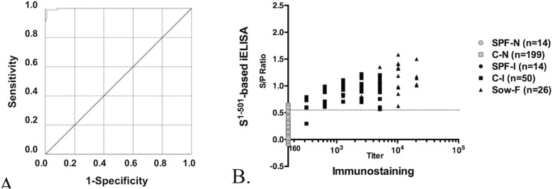
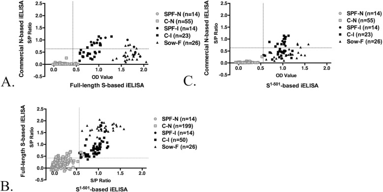
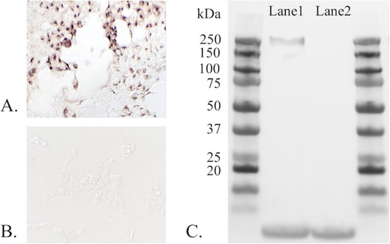
Background: Since 2010, outbreaks of genotype 2 (G2) porcine epidemic diarrhea virus (PEDV) have caused high mortality in neonatal piglets and have had devastating impacts on the swine industry in many countries. A reliable serological assay for evaluating the PEDV-specific humoral and mucosal immune response is important for disease survey, monitoring the efficacy of immunization, and designing strategies for the prevention and control of PED. Two PEDV spike (S) glycoprotein-based indirect enzyme-linked immunosorbent assays (ELISAs) were developed using G2b PEDV-Pintung 52 (PEDV-PT) trimeric full-length S and truncated S1-501 proteins derived from the human embryonic kidney (HEK)-293 cell expression system. The truncated S1-501 protein was selected from a superior expressed stable cell line. The sensitivity and specificity of these two ELISAs were compared to immunostaining of G2b PEDV-PT infected cells and to a commercial nucleocapsid (N)-based indirect ELISA kit using a panel of PEDV negative and hyperimmune sera.

Results: The commercial N-based ELISA exhibited a sensitivity of 37%, a specificity of 100%, and a fair agreement (kappa = 0.37) with the immunostaining result. In comparison, the full-length S-based ELISA showed a sensitivity of 97.8%, a specificity of 94%, and an almost perfect agreement (kappa = 0.90) with the immunostaining result. Interestingly, the S1-501-based ELISA had even higher sensitivity of 98.9% and specificity of 99.1%, and an almost perfect agreement (kappa = 0.97) with the immunostaining result. A fair agreement (kappa< 0.4) was seen between the commercial N-based ELISA and either of our S-based ELISAs. However, the results of the full-length S-based ELISA shared an almost perfect agreement (kappa = 0.92) with that of S1-501-based ELISA.

Conclusions: Both full-length S-based and S1-501-based ELISAs exhibit high sensitivity and high specificity for detecting antibodies against PEDVs. Considering the high protein yield and cost-effectiveness, the S1-501-based ELISA could be used as a reliable, sensitive, specific, and economic serological test for PEDV.

Keywords: Human embryonic kidney (HEK)-293 mammalian cell expression system; Indirect ELISA; Porcine epidemic diarrhea (PED); Serology; Spike protein specific ELISA.


Conflict of interest statement

The authors declare that they have no competing interests.

Figures

Fig. 1
Fig. 1
Representative images of immunostaining of serum samples against G2b PEDV-PT infected Vero cells. Vero cells were seeded onto 96-well plates and grown overnight until reaching 90% confluency. The Vero cells were challenged with 1000 TCID50/mL of G2b PEDV-PT-P6 diluted in post-inoculation (PI) medium. Vero cells with syncytial cells, the cytopathic effect (CPE) of PEDV, were fixed with acetone after 24 h of incubation. Each serum sample from different sources was serially diluted from 160-fold to 20,480-fold, then added to the plates and incubated for 1 h. Anti-pig IgG antibodies conjugated with horseradish peroxidase (HRP) or fluorescence were used to probe serum IgG on the syncytial cells. A positive signal was found in the Vero cells showing CPE. a The representative “positive” result of immunostaining using the serum sample from the C-I group at a 640-fold dilution. b The representative “negative” result of immunostaining using the serum sample from the C-N group at a 640-fold dilution. The bars represent the scale of 100 μm
Fig. 2
Fig. 2
Distribution of the results of the immunostaining and commercial N-based ELISA. The immunostaining titers of each serum sample were presented as logarithms with the base 10. Serum samples which were negative for the immunostaining are denoted by 0. The results of the commercial N-based ELISA kit were presented by the sample to positive ratio (S/P ratio). S/P ratio = [(sample OD – mean OD of negative controls) / (mean OD of positive controls – mean OD of negative controls)]. The dotted line represents the threshold of an S/P ratio of 0.6 of the commercial N-based ELISA, as per the manufacturer’s instructions. C-N (with grey square icon): conventional PEDV naïve pigs; C-I (with black square icon): experimental PEDV inoculated conventional pigs; SPF-N (with grey round icon): specific pathogen free PEDV naïve pigs; SPF-I (with black round icon): experimental PEDV inoculated SPF pigs; Sow-F (with black triangle icon): PEDV-feedback sows
Fig. 3
Fig. 3
Receiver operating characteristic (ROC) analysis and distribution of the results of the immunostaining and full-length S-based ELISA. a The ROC analysis of the full-length S-based ELISA. The sensitivity and specificity were calculated using negative (n = 213) and positive (n = 90) serum samples. The x axis shows 1-specificity and the y axis shows sensitivity. b The immunostaining titers of immunofluorescence assay of each serum sample are presented as a logarithm with base 10. Serum samples that were negative for the immunostaining are denoted by 0. The results of the full-length S-based ELISA are presented as the S/P ratio. The dotted line represents the cut-off value of an S/P ratio of 0.432 as determined by the ROC analysis. C-N (with grey square icon): conventional PEDV naïve pigs; C-I (with black square icon): experimental PEDV inoculated conventional pigs; SPF-N (with grey round icon): specific pathogen free PEDV naïve pigs; SPF-I (with black round icon): experimental PEDV inoculated SPF pigs; Sow-F (with black triangle icon): PEDV-feedback sows
Fig. 4
Fig. 4
ROC analysis and the distribution of the results of the immunostaining and S1–501-based ELISA. a ROC analysis of the S1–501-based ELISA. The sensitivity and specificity were calculated using negative (n = 213) and positive (n = 90) serum samples. The x axis shows 1-specificity and the y axis shows sensitivity. b The immunostaining titers of immunofluorescence assay of each serum sample are presented as a logarithm with base 10. The serum samples that were negative for the immunostaining are denoted by 0. The results of the S1–501-based ELISA are presented as the S/P ratio. The dotted line represents the cut-off value of an S/P ratio of 0.573 as determined by the ROC analysis. C-N (with grey square icon): conventional PEDV naïve pigs; C-I (with black square icon): experimental PEDV inoculated conventional pigs; SPF-N (with grey round icon): specific pathogen free PEDV naïve pigs; SPF-I (with black round icon): experimental PEDV inoculated SPF pigs; Sow-F (with black triangle icon): PEDV-feedback sows
Fig. 5
Fig. 5
Comparison between commercial N-based, full-length S-based, and S1–501-based ELISAs. a The distribution of the results of the full-length S-based and commercial N-based ELISAs. The vertical dotted line represents the cut-off value of the full-length-based ELISA. The horizontal dotted line represents the threshold of the commercial N-based ELISAs, as suggested by the manufacturer. b Distribution of the results of S1–501-based and full-length S-based ELISAs. The vertical dotted line represents the cut-off value of the S1–501-based ELISA. The horizontal line represents the cut-off value of the S/P ratio of the full-length-based ELISA. c The distribution of the results of S1–501-based and commercial N-based ELISAs. The vertical dotted line represents the cut-off value of the S1–501-based ELISA. The horizontal dotted line represents the threshold of the commercial N-based ELISAs, as suggested by the manufacturer. C-N (with grey square icon): conventional PEDV naïve pigs; C-I (with black square icon): experimental PEDV inoculated conventional pigs; SPF-N (with grey round icon): specific pathogen free PEDV naïve pigs; SPF-I (with black round icon): experimental PEDV inoculated SPF pigs; Sow-F (with black triangle icon): PEDV-feedback sows
Fig. 6
Fig. 6
Detection of PEDV S-specific IgA from colostrum using the full-length S-based and S1–501-based ELISAs. Colostrum samples from five seronegative SPF sows (n = 5, SPF group) and twenty-three feedback conventional sows (n = 23, Conventional group) were included. The PEDV S-specific IgA was detected from the colostrum using either full-length S-based and S1–501-based ELISAs. The results are presented as the S/P ratio. The round icon represents the result of SPF group; the square icon represents the result of Conventional group. The error bars indicate the mean S/P ratio ± SD
Fig. 7
Fig. 7
Expression and characterization of G1b PEDV S protein by ICC staining and western blotting. a ICC staining of G1b PEDV S-expressing HEK cells using anti-V5 tag antibody. The cells were fixed on the plates using acetone, probed using 1000-fold diluted anti-V5 tag antibody, and detected using anti-mouse IgG antibody conjugated with HRP. The coloration procedure was carried out using the DAB system. b The negative control of the ICC staining of non-transfected HEK cells. The cells were probed with anti-V5 tag antibody following by the anti-mouse IgG antibody conjugated with HRP to distinguish the non-specific signals. c The molecular weight of the purified G1b PEDV S protein (Lane 1) was estimated by western blotting. The purified G1b PEDV S protein was denatured in NuPAGE® LDS sample buffer containing the NuPAGE® reducing agent and boiled at 95 °C for 5 min, then, separated by SDS-PAGE, transferred onto the PVDF membrane, and stained with anti-V5 tag antibody. The HEK cell lysate was used as the negative control (Lane 2). The ladder is shown in kilodalton (kDa)

References

    1. Jung K, Saif LJ. Porcine epidemic diarrhea virus infection: etiology, epidemiology, pathogenesis and immunoprophylaxis. Vet J. 2015;204(2):134–143. doi: 10.1016/j.tvjl.2015.02.017. - DOI - PMC - PubMed
    1. Jung K, Annamalai T, Lu Z, Saif LJ. Comparative pathogenesis of US porcine epidemic diarrhea virus (PEDV) strain PC21A in conventional 9-day-old nursing piglets vs. 26-day-old weaned pigs. Vet Microbiol. 2015;178(1):31–40. doi: 10.1016/j.vetmic.2015.04.022. - DOI - PMC - PubMed
    1. Wang Q, Vlasova AN, Kenney SP, Saif LJ. Emerging and re-emerging coronaviruses in pigs. Curr Opin Virol. 2019;34:39–49. doi: 10.1016/j.coviro.2018.12.001. - DOI - PMC - PubMed
    1. Song D, Moon H, Kang B. Porcine epidemic diarrhea: a review of current epidemiology and available vaccines. Clin Exp Vaccine Res. 2015;4(2):166–176. doi: 10.7774/cevr.2015.4.2.166. - DOI - PMC - PubMed
    1. Lai MM, Cavanagh D. The molecular biology of coronaviruses. Adv Virus Res. 1997;48:1–100. - PMC - PubMed

MeSH terms

Substances

LinkOut - more resources