Review
. 2019 Nov 27;17(1):156.
doi: 10.1186/s12964-019-0461-0.

TAM receptors, Phosphatidylserine, inflammation, and Cancer

Affiliations
Review

TAM receptors, Phosphatidylserine, inflammation, and Cancer

Tal Burstyn-Cohen et al. Cell Commun Signal. .

Abstract

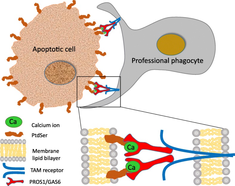
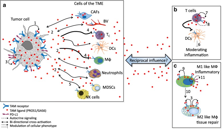
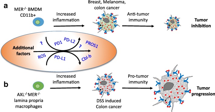
The numerous and diverse biological roles of Phosphatidylserine (PtdSer) are featured in this special issue. This review will focus on PtdSer as a cofactor required for stimulating TYRO3, AXL and MERTK - comprising the TAM family of receptor tyrosine kinases by their ligands Protein S (PROS1) and growth-arrest-specific 6 (GAS6) in inflammation and cancer. As PtdSer binding to TAMs is a requirement for their activation, the biological repertoire of PtdSer is now recognized to be broadened to include functions performed by TAMs. These include key homeostatic roles necessary for preserving a healthy steady state in different tissues, controlling inflammation and further additional roles in diseased states and cancer. The impact of PtdSer on inflammation and cancer through TAM signaling is a highly dynamic field of research. This review will focus on PtdSer as a necessary component of the TAM receptor-ligand complex, and for maximal TAM signaling. In particular, interactions between tumor cells and their immediate environment - the tumor microenvironment (TME) are highlighted, as both cancer cells and TME express TAMs and secrete their ligands, providing a nexus for a multifold of cross-signaling pathways which affects both immune cells and inflammation as well as tumor cell biology and growth. Here, we will highlight the current and emerging knowledge on the implications of PtdSer on TAM signaling, inflammation and cancer.

Keywords: Cancer; GAS6; Inflammation; PROS1; Phosphatidylserine; Protein S; PtdSer; TAM receptors.


Conflict of interest statement

The authors declare that they have no competing interests.

Figures

Fig. 1
Fig. 1
TAM - PtdSer association and the uptake of apoptotic cells. Apoptotic cells (ACs) externalize phosphatidylserine (PtdSer) which is bound by the TAM ligands GAS6 and PROS1. This binding occurs at the GLA domain, present at the amino terminus of the ligands, and is enhanced by the presence of calcium ions, depicted by green ovals. The carboxyl terminal of PROS1 and GAS6 binds to the extracellular domains of the TAM receptors, present on professional phagocytes such as retinal pigment epithelium, Sertoli cells, osteoclasts, macrophages and DCs. TAM receptor activation is optimal in the presence of both ligands and PtdSer. By binding PtdSer on one side and to TAM receptors on the phagocytic cell, PROS1 and GAS6 function as bridging molecules physically linking the phagocyte to the engulfed PtdSer-decorated moiety. In case of macrophage and dendritic cell phagocytes, AC uptake and TAM activation also results in shutting inflammatory signaling and cytokine secretion. Abbreviations: Ca – Calcium ion; PtdSer – phosphatidylserine; TAM – TYRO3, AXL, MERTK, PROS1 – protein S; GAS6 – growth arrest specific 6
Fig. 2
Fig. 2
TAM - PtdSer interactions in the tumor microenvironment. Autocrine and paracrine cross-signaling through PtdSer-TAM in a tumor setting. (a) Both cancer cells and the different TME cellular compartments express TAM receptors and secrete PROS1 and GAS6. The abundance of PtdSer enables potent autocrine (1) and / or paracrine (2) activation of TAM receptors expressed by tumor cells, resulting in augmented aggressiveness, also by inducing expression of the immune evasion/checkpoint molecules PD-L1 on cancer cells (3, [66]). Tumor-derived TAM ligands suppresses macrophage and T cell infiltration (4, [69, 70]. Similarly, the antitumor cytotoxicity of NK cells is suppressed by TAM receptor expression (5, [33]. (b) PtdSer-TAM signaling plays a role in immune cells, where they dampen inflammation, as described for the interactions between T and dendritic cells (6, [23]). Within T cells, opposing roles for TAM signaling report MERTK-dependent signaling to suppresses T cell activation and promote immune evasion through induction of PD-1 expression (7, [71]), but also to provide co-stimulatory functions (8, [72]). (c) In the case of macrophages, reports indicate that PstSer-TAM signaling is chiefly anti-inflammatory due to autocrine signaling within M1 and M2-like macrophages (9, [73]), and shifts M1-like pro-inflammatory macrophages towards the anti-inflammatory M2-like state (10, [74]), but also promotes anti-immunity through PD-L1 and PD-L2 expression (11, [71]). Altogether, although PtdSer-TAM signaling may result in opposing outcomes, the net effect of all interactions contributes to the generation of tumors with superior tumorigenic characteristics, within a more permissive environment. See text for details. Abbreviations: CAFs – cancer associated fibroblasts; BV – blood vessel; Tc – T cells; NK – natural killer, MDSCs – myeloid derived suppressor cells; DCs – dendritic cells; MФ – macrophage; TME – tumor microenvironment
Fig. 3
Fig. 3
Inhibition of PtdSer-TAM signaling in tumor-immune interactions leads to elevated inflammation but may differentially affect tumor growth. Bone-marrow derived macrophages (BMDMs) differentially influence tumor progression in different cancer models. (a) Inhibition of MERTK in CD11b + BMDMs resulted in elevated inflammation, which conveyed anti-tumor immunity resulting in inhibited growth of breast, melanoma and MC38 colon cancer tumors [69]. (b) By contrast to (a), in a DSS-induced model of colon cancer, the dual inhibition of AXL and MERTK in BMDMs had no effect on tumor progression [76]. Instead, AXL and MERK inhibition in F4/80+; CD11b + lamina propria macrophages conveyed pro-tumor immunity, which promoted cancer progression. These data demonstrate that while inhibition of TAM signaling in macrophages led to inflammation in both cases, opposing effects were imparted on tumor growth, highlighting the complex liaisons between immune and tumor cells through inflammation. Such complexity is likely to be mediated by additional factors, some of which function by immune-modulation, others are yet to be revealed (depicted in the oval). See discussion in the main text

References

    1. Lemke G. Biology of the TAM receptors. Cold Spring Harb Perspect Biol. 2013;5:a009076. doi: 10.1101/cshperspect.a009076. - DOI - PMC - PubMed
    1. Rothlin CV, Carrera-Silva EA, Bosurgi L, Ghosh S. TAM receptor signaling in immune homeostasis. Annu Rev Immunol. 2015;33:355–391. doi: 10.1146/annurev-immunol-032414-112103. - DOI - PMC - PubMed
    1. Burstyn-Cohen T. TAM receptor signaling in development. The International journal of developmental biology. 2017;61:215–224. doi: 10.1387/ijdb.160285tb. - DOI - PubMed
    1. Zelentsova-Levitskyi K, Talmi Z, Abboud-Jarrous G, Capucha T, Sapir T, Burstyn-Cohen T. Protein S negatively regulates neural stem cell self-renewal through Bmi-1 signaling. Front Mol Neurosci. 2017;10:124. - PMC - PubMed
    1. Zelentsova K, Talmi Z, Abboud-Jarrous G, Sapir T, Capucha T, Nassar M, Burstyn-Cohen T. Protein S regulates neural stem cell quiescence and neurogenesis. Stem Cells. 2016;35:679–693. doi: 10.1002/stem.2522. - DOI - PubMed

Publication types

Substances