

Morphological characteristics of different types of distal radius die-punch fractures based on three-column theory
- PMID: 31775810
- PMCID: PMC6882215
- DOI: 10.1186/s13018-019-1453-x
Morphological characteristics of different types of distal radius die-punch fractures based on three-column theory
Abstract
Objective: The aim of this study is to investigate the morphological characteristics of distal radius die-punch fracture (DRDPF) with different types, based on the three-column theory.
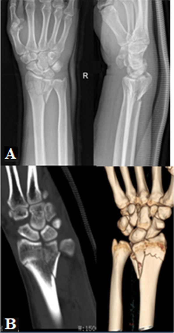
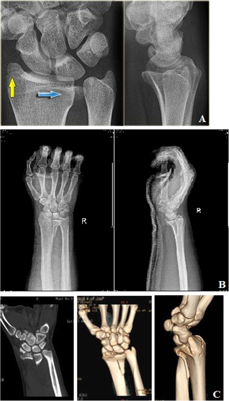
Methods: The imaging data of 560 patients diagnosed with DRDPF were reviewed and divided into single-column, double-column, or three-column DRDPF according to the three-column theory, and the types, case distribution of DRDPF, and inter- and intra-agreement of classification were further analyzed.
Results: There were 65 cases of single-column DRDPF, 406 cases of double-column DRDPF, and 89 cases of three-column DRDPF. Among the single-column DRDPF, there were three cases of volar, 13 cases of dorsal, 14 cases of split, and 35 cases of collapse type fractures. Among the radius column fracture, there were 130 cases of metaphseal,155 cases of articular surface, and 210 cases of combined type. The inter-observer Kappa coefficient was 0.877-0.937, and the intra-observer kappa was 0.916-0.959, showing high agreement. At the 12th month's follow-up, according to the Gartland-Werley score system for the functionary recovery of the wrist and hand, 519 cases (92.68%) of the patients ranked excellent or good, and 41 cases (7.32%) ranked fair. All the cases were fair results, and the intermediate column of the distal radius was collapse type fractures, showing significant difference between the collapse type and other types (χ2 = 23.460, P = 0.000). The excellent and good rate in the single-, double-, and three-column DRDPFs were 93.85%, 92.16%, and 91.01%, respectively (χ2 = 0.018, P = 0.991).
Conclusion: Due to the difference of the nature and energy of the forces, the position of wrist, and the bone quality of the patients at the moment of the injury, the loading forces transmitted to the intermediate column of the distal radius could result in different types of DRDPF. The classification method in this study included all types of DRDPF, indicating the mechanism, affected sites, and the morphological characteristics of DRDPF with high consistency, which hopefully could provide insight into the treatment and prognosis of DRDPF patients.
Keywords: Die-punch fracture; Distal radius; Three-column theory.
Conflict of interest statement
The authors declare that they have no competing interests.
Figures


References
MeSH terms
LinkOut - more resources
Full Text Sources
Medical
