

MFAP5 facilitates the aggressiveness of intrahepatic Cholangiocarcinoma by activating the Notch1 signaling pathway
- PMID: 31775892
- PMCID: PMC6882185
- DOI: 10.1186/s13046-019-1477-4
MFAP5 facilitates the aggressiveness of intrahepatic Cholangiocarcinoma by activating the Notch1 signaling pathway
Abstract
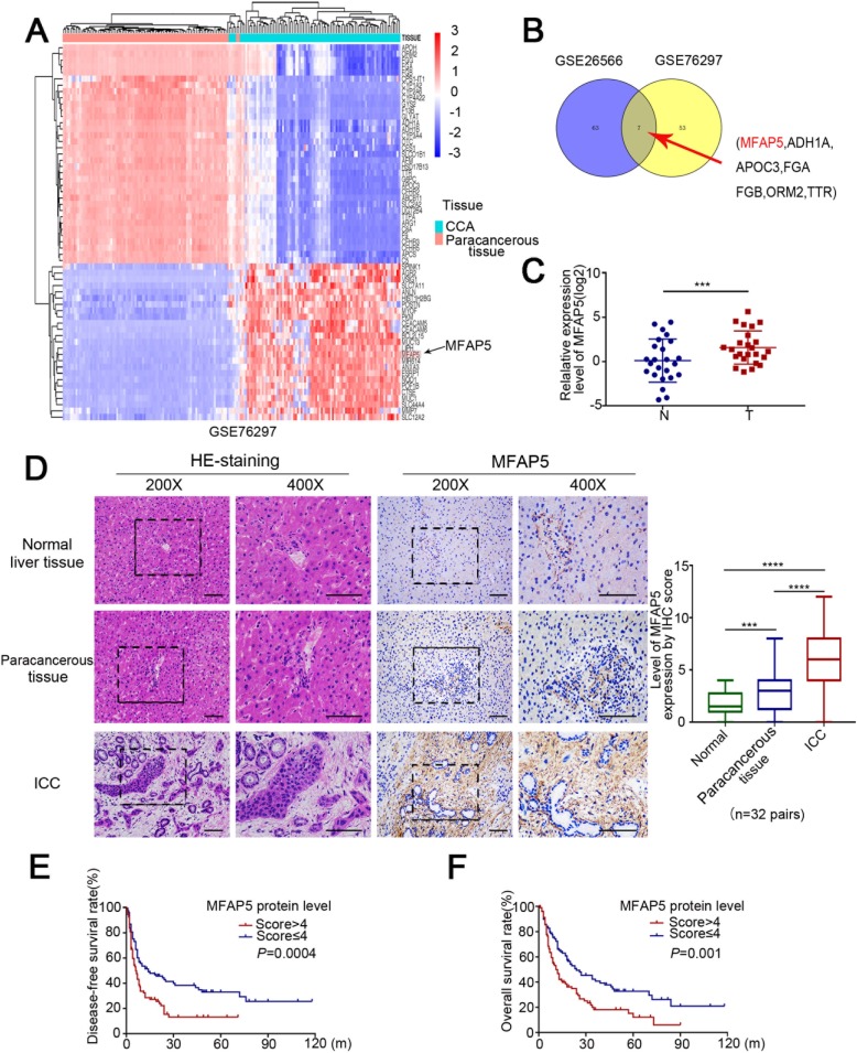
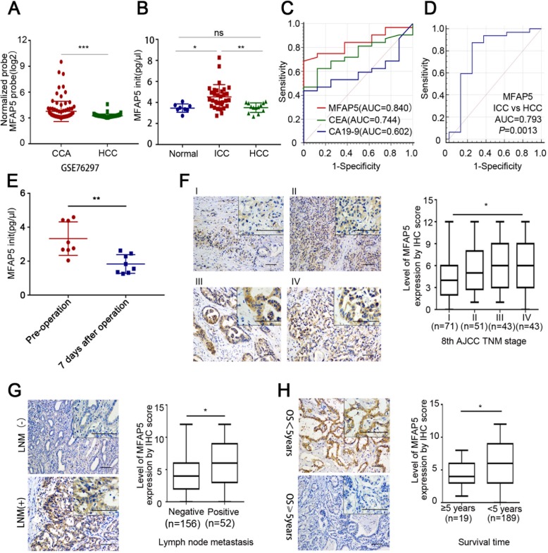
Background: Intrahepatic cholangiocarcinoma (ICC) is the second most common primary liver cancer. The dismal outcome of ICC patients is due to lack of early diagnosis, the aggressive biological behavior of ICC and the lack of effective therapeutic options. Early diagnosis and prognosis of ICC by non-invasive methods would be helpful in providing valuable information and developing effective treatment strategies.
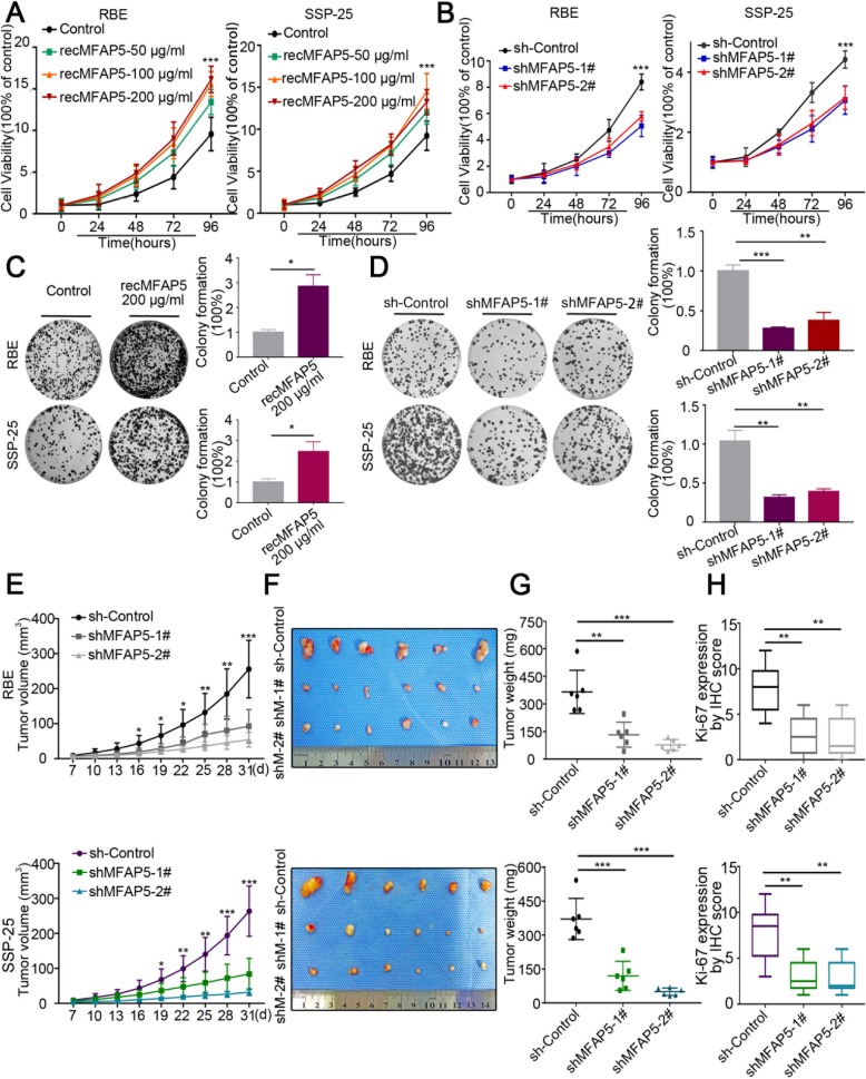
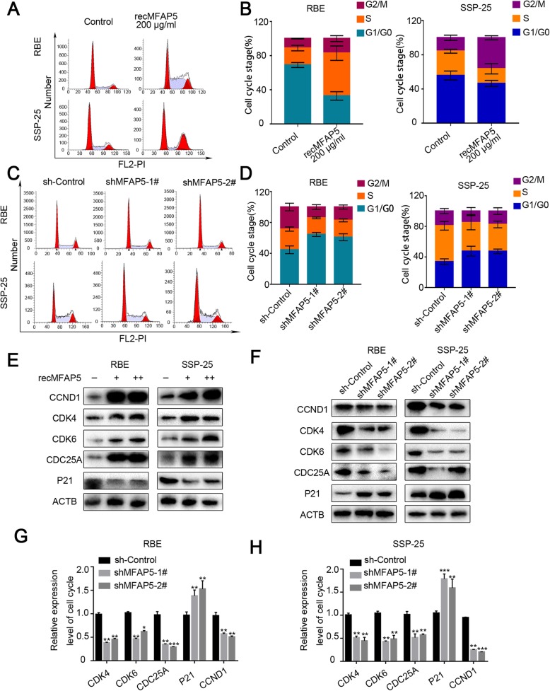
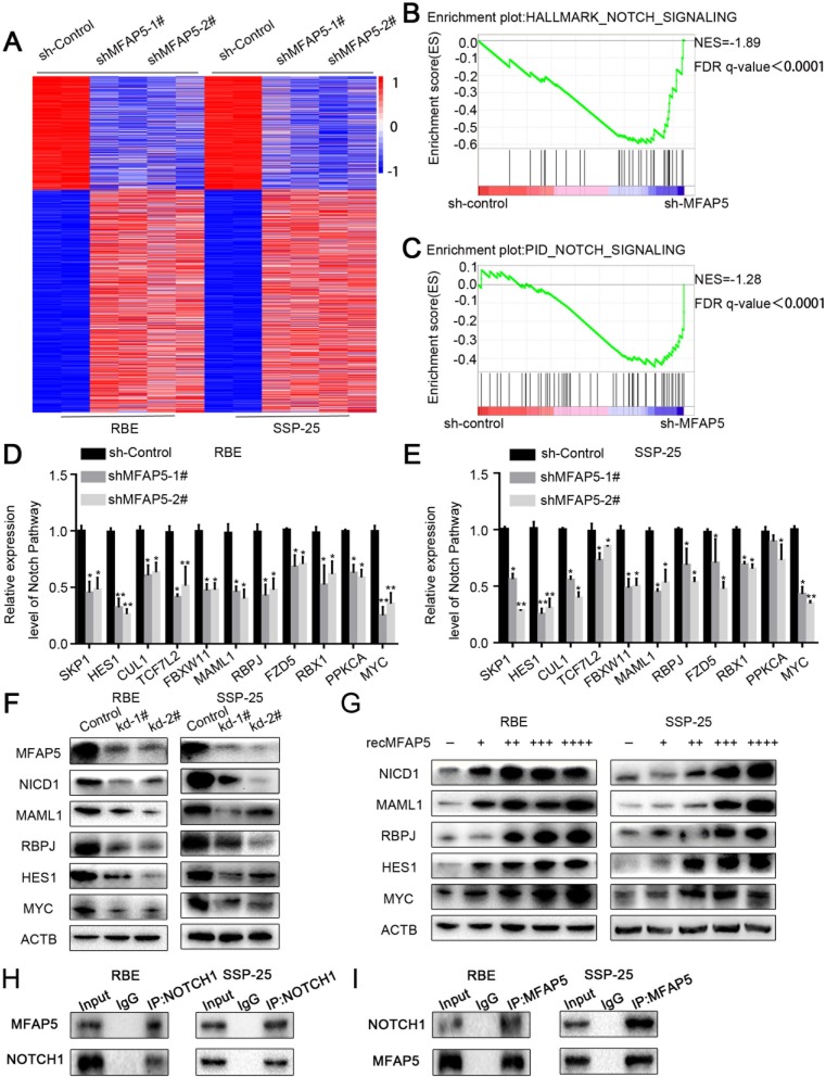
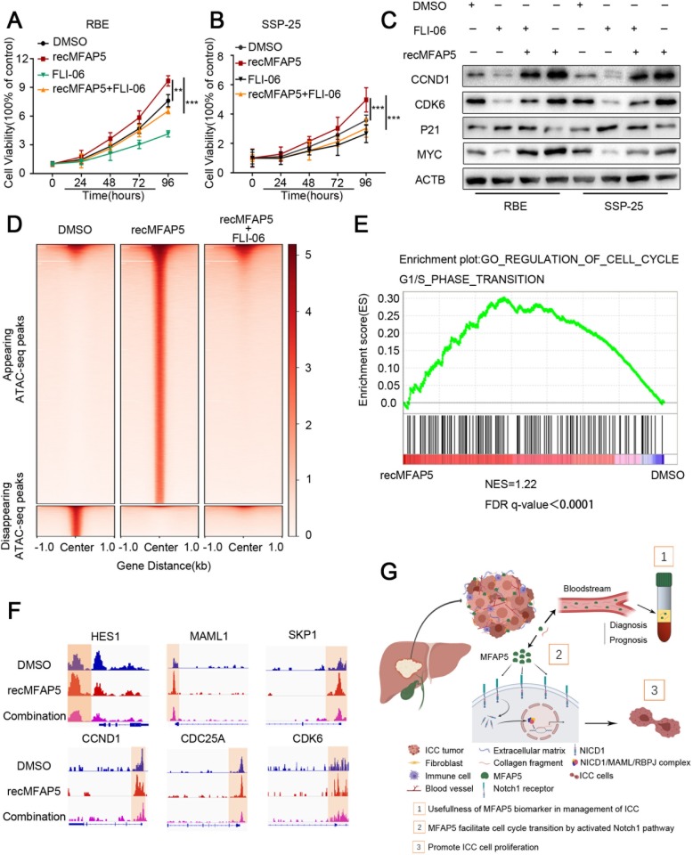
Methods: Expression of microfibrillar-associated protein 5 (MFAP5) in the serum of ICC patients was detected by ELISA. Human ICC specimens were immunostained by MFAP5 antibodies. The growth rate of human ICC cell lines treated with MFAP5 or MFAP5 shRNAs was examined by CCK8 and colony formation assays. Cell cycle analysis was performed with PI staining. The effect of MFAP5 inhibition was assessed by xenograft models in nude mice. RNA-seq and ATAC-seq analyses were used to dissect the molecular mechanism by which MFAP5 promoted ICC aggressiveness.
Results: We identified MFAP5 as a biomarker for the diagnosis and prognosis of ICC. Upregulated MFAP5 is a common feature in aggressive ICC patients' tissues. Importantly, MFAP5 level in the serum of ICC patients and healthy individuals showed significant differential expression profiles. Furthermore, we showed that MFAP5 promoted ICC cell growth and G1 to S-phase transition. Using RNA-seq expression and ATAC-seq chromatin accessibility profiling of ICC cells with suppressed MFAP5 secretion, we showed that MFAP5 regulated the expression of genes involved in the Notch1 signaling pathway. Furthermore, FLI-06, a Notch signaling inhibitor, completely abolished the MFAP5-dependent transcriptional programs.
Conclusions: Raised MFAP5 serum level is useful for differentiating ICC patients from healthy individuals, and could be helpful in ICC diagnosis, prognosis and therapies.
Keywords: Cancer biomarkers; Cell cycle arrest; Intrahepatic cholangiocarcinoma; MFAP5; NOTCH.
Conflict of interest statement
The authors declare that they have no competing interests.
Figures
Similar articles
-
Notch1 Drives the Formation and Proliferation of Intrahepatic Cholangiocarcinoma.Curr Med Sci. 2019 Dec;39(6):929-937. doi: 10.1007/s11596-019-2125-0. Epub 2019 Dec 16. Curr Med Sci. 2019. PMID: 31845224
-
The PKHD1 gene inhibits tumor proliferation and invasion in intrahepatic cholangiocarcinoma by activating the Notch pathway.Int J Med Sci. 2024 Oct 14;21(14):2655-2663. doi: 10.7150/ijms.95964. eCollection 2024. Int J Med Sci. 2024. PMID: 39512694 Free PMC article.
-
Notch1 is overexpressed in human intrahepatic cholangiocarcinoma and is associated with its proliferation, invasiveness and sensitivity to 5-fluorouracil in vitro.Oncol Rep. 2014 Jun;31(6):2515-24. doi: 10.3892/or.2014.3123. Epub 2014 Apr 2. Oncol Rep. 2014. PMID: 24700253
-
Multiple cellular origins and molecular evolution of intrahepatic cholangiocarcinoma.Cancer Lett. 2016 Sep 1;379(2):253-61. doi: 10.1016/j.canlet.2016.02.038. Epub 2016 Mar 2. Cancer Lett. 2016. PMID: 26940139 Review.
-
The role of microRNAs in intrahepatic cholangiocarcinoma.J Cell Mol Med. 2017 Jan;21(1):177-184. doi: 10.1111/jcmm.12951. Epub 2016 Sep 13. J Cell Mol Med. 2017. PMID: 27619971 Free PMC article. Review.
Cited by
-
Atractylodin and β-eudesmol from Atractylodes lancea (Thunb.) DC. Inhibit Cholangiocarcinoma Cell Proliferation by Downregulating the Notch Signaling Pathway.Asian Pac J Cancer Prev. 2023 Feb 1;24(2):551-558. doi: 10.31557/APJCP.2023.24.2.551. Asian Pac J Cancer Prev. 2023. PMID: 36853304 Free PMC article.
-
Microfibril-associated protein 5 and the regulation of skin scar formation.Sci Rep. 2023 May 30;13(1):8728. doi: 10.1038/s41598-023-35558-x. Sci Rep. 2023. PMID: 37253753 Free PMC article.
-
MFAP5 Strengthened the Stem Cell Features of Non-small Cell Lung Cancer Cells by Regulating the FBW/Sox9 Axis.Curr Pharm Biotechnol. 2025;26(2):235-245. doi: 10.2174/0113892010259632240213091136. Curr Pharm Biotechnol. 2025. PMID: 38415489
-
Notch signaling in the pathogenesis, progression and identification of potential targets for cholangiocarcinoma (Review).Mol Clin Oncol. 2022 Mar;16(3):66. doi: 10.3892/mco.2022.2499. Epub 2022 Jan 19. Mol Clin Oncol. 2022. PMID: 35154706 Free PMC article. Review.
-
MFAP5 inhibits the malignant progression of endometrial cancer cells in vitro.Open Life Sci. 2024 Dec 31;19(1):20220990. doi: 10.1515/biol-2022-0990. eCollection 2024. Open Life Sci. 2024. PMID: 39759103 Free PMC article.
References
MeSH terms
Substances
Grants and funding
- 81772522/National Natural Science Foundation of China
- 81472261/National Natural Science Foundation of China
- 81572766/National Natural Science Foundation of China
- 31771630/National Natural Science Foundation of China
- 2016A030313238/Natural Science Foundation of Guangdong Province
- 2017A030312009/Natural Science Foundation of Guangdong Province
- 2019A1515010686/Natural Science Foundation of Guangdong Province
- 201604020044/Science and Technology Development Projects of Guangzhou, Guangdong, China
- 2016ZT06S029/Guangdong Innovative and Entrepreneurial Research Team Program
- 2016M602588/Postdoctoral Research Foundation of China
- 2108M643325/Postdoctoral Research Foundation of China
- 2108M643327/Postdoctoral Research Foundation of China
- KY20160309/Shenzhen Science Foundation
LinkOut - more resources
Full Text Sources
Medical
Research Materials
Miscellaneous
