. 2019 Dec 17;116(51):25891-25899.
doi: 10.1073/pnas.1910334116. Epub 2019 Nov 27.

Combined HIV-1 sequence and integration site analysis informs viral dynamics and allows reconstruction of replicating viral ancestors

Affiliations

Combined HIV-1 sequence and integration site analysis informs viral dynamics and allows reconstruction of replicating viral ancestors

Sean C Patro et al. Proc Natl Acad Sci U S A. .

Abstract

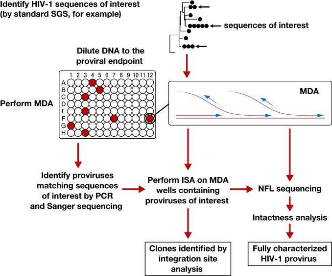
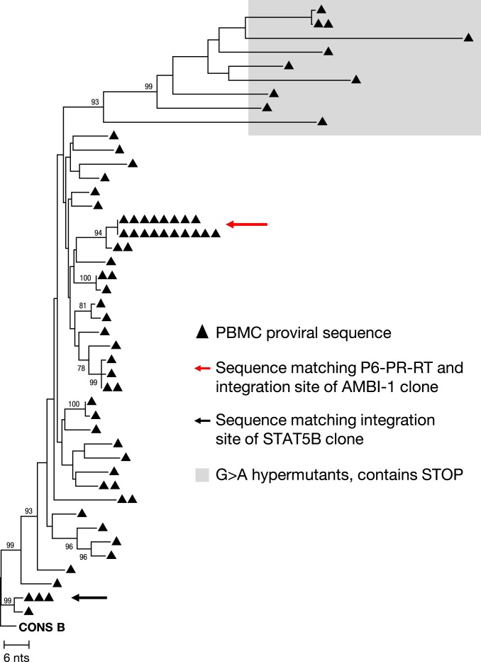
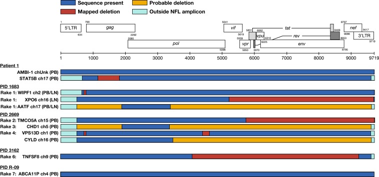
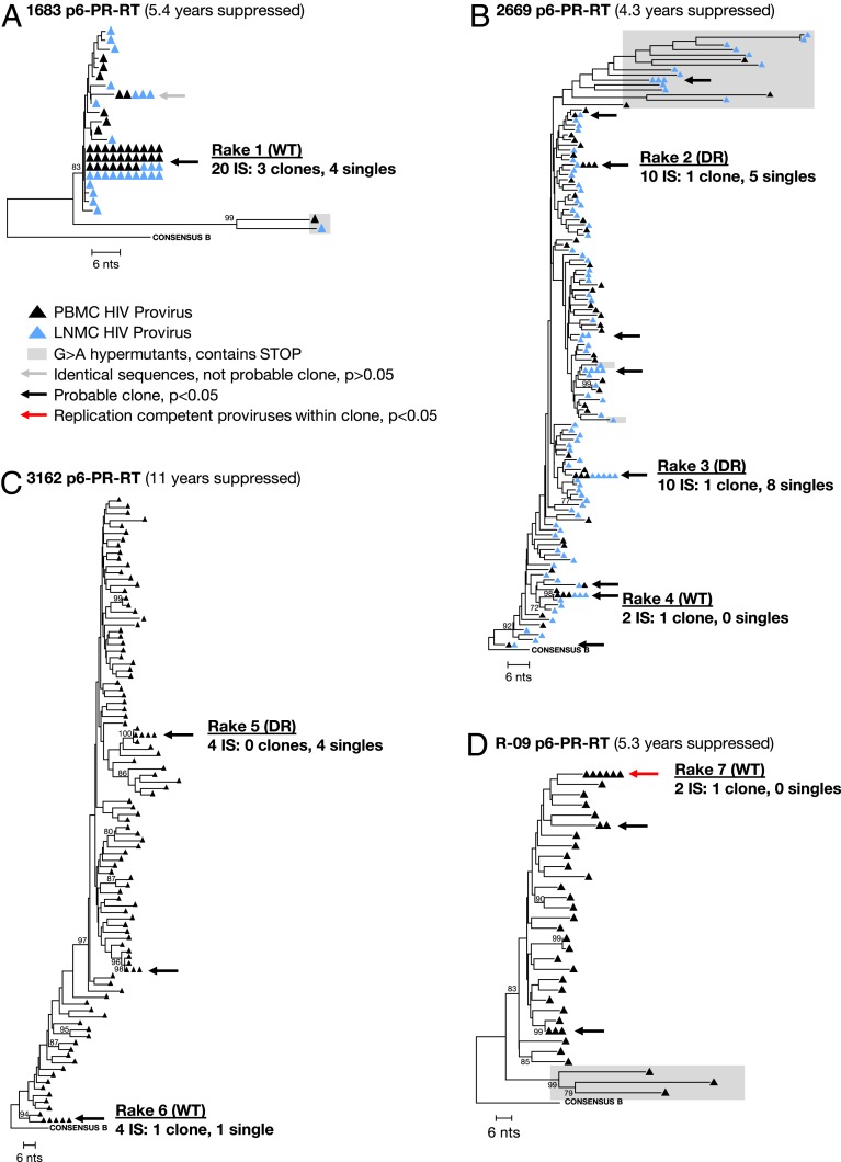
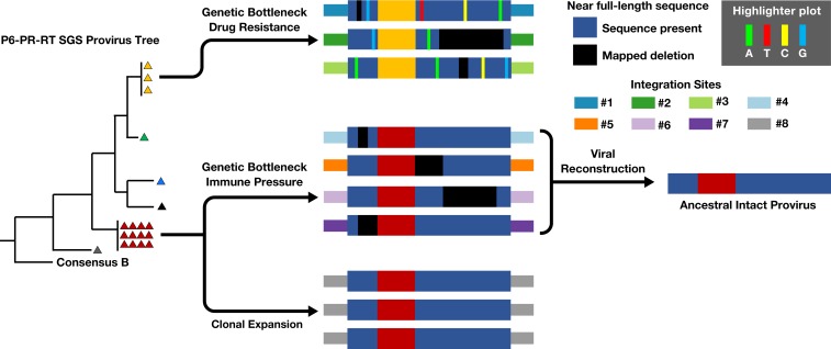
Understanding HIV-1 persistence despite antiretroviral therapy (ART) is of paramount importance. Both single-genome sequencing (SGS) and integration site analysis (ISA) provide useful information regarding the structure of persistent HIV DNA populations; however, until recently, there was no way to link integration sites to their cognate proviral sequences. Here, we used multiple-displacement amplification (MDA) of cellular DNA diluted to a proviral endpoint to obtain full-length proviral sequences and their corresponding sites of integration. We applied this method to lymph node and peripheral blood mononuclear cells from 5 ART-treated donors to determine whether groups of identical subgenomic sequences in the 2 compartments are the result of clonal expansion of infected cells or a viral genetic bottleneck. We found that identical proviral sequences can result from both cellular expansion and viral genetic bottlenecks occurring prior to ART initiation and following ART failure. We identified an expanded T cell clone carrying an intact provirus that matched a variant previously detected by viral outgrowth assays and expanded clones with wild-type and drug-resistant defective proviruses. We also found 2 clones from 1 donor that carried identical proviruses except for nonoverlapping deletions, from which we could infer the sequence of the intact parental virus. Thus, MDA-SGS can be used for "viral reconstruction" to better understand intrapatient HIV-1 evolution and to determine the clonality and structure of proviruses within expanded clones, including those with drug-resistant mutations. Importantly, we demonstrate that identical sequences observed by standard SGS are not always sufficient to establish proviral clonality.

Keywords: HIV persistence; integration site analysis; proviral structure.


Conflict of interest statement

Competing interest statement: J.W.M. is a consultant to Gilead Sciences, Merck Research Laboratories, Janssen Pharmaceuticals, and AccelevirDx, and a share option holder of Co-Crystal, Inc. B.F.K. and J.A.H. are co-authors on an October 2015 article. The remaining authors have no potential conflicts.

Figures

Fig. 1.
Fig. 1.
Workflow for MDA-SGS. Extracted DNA is aliquoted so that individual proviruses, along with background genomic DNA, can be independently amplified ∼1,000-fold by MDA. This amplification allows the same provirus to be identified by SGS (–3), analyzed by ISA (integration sites analysis) (1, 10, 22) and NFL (near–full-length) sequencing, followed by further downstream analyses, if needed. Infected cell clones are identified by finding identical integration sites across multiple MDA wells.
Fig. 2.
Fig. 2.
Neighbor-joining phylogenetic tree of P6–PR–RT MDA-SGS on effector memory T cells from patient 1. The red arrow indicates sequences matching P6–PR–RT of the provirus in the AMBI-1 clone, a provirus resulting in virus continuously recovered in plasma during ART and shown to be replication competent (1, 10). The black arrow indicates group of identical sequences with integration site matching the STAT5B expanded clone reported in Maldarelli et al. (1). Bootstrap values are shown for nodes greater than 70% confidence.
Fig. 3.
Fig. 3.
Near–full-length (NFL) proviral structures. Structures of proviruses within cell clones aligned to the HXB2 reference sequence (adapted from Los Alamos National Laboratory). Corresponding rake ID (Fig. 4), integration site (gene/nearest gene, chromosome), and cell source (PB, LN) are given. A mapped deletion (red box) denotes sequence obtained spanning the deletion. Probable deletion (yellow box) denotes failure at either amplification or sequencing. All proviruses detected were shown to be defective except for the AMBI-1 provirus and the newly identified ABCA11P provirus in R-09. LN, lymph node; PB, peripheral blood.
Fig. 4.
Fig. 4.
Neighbor-joining trees of viral sequences obtained from donors on ART. SGS of P6–PR–RT region of viral DNA from SCOPE donors 1683 (A), 2669 (B), 3162 (C), and Pitt donor R-09 (D). Average pairwise distance: 0.2%, 1.6%, 2.7%, and 1.9%, respectively. The gray arrow indicates a group that had fewer than the number of identical sequences expected by chance (P > 0.05); the black arrows indicate groups with more than the expected number of identical sequences, and the red arrow indicates groups that matched variants that grew out in VOA. Groups that were investigated for clonality are labeled rake #1 to #7. Within each group, the number of total integration sites, confirmed expanded clones, and single integration sites are given. Integration site (IS), wild type (WT), and drug-resistance mutations (DR). Bootstrap values are shown for nodes with greater than 70% confidence.
Fig. 5.
Fig. 5.
Origin of Identical P6–PR–RT sequences. Identical subgenomic P6–PR–RT sequences observed during ART can be originate from multiple mechanisms including (Top) genetic bottleneck of multiple viruses during selection of drug resistance, (Middle) immune pressure (i.e., CTL/antibody) driving persistence of select proviruses, and (Bottom) clonal expansion of cells containing a provirus with identical integration site and sequence. Hypothetical integration sites and proviral deletions are shown. Reconstruction of the replication-competent ancestor resulting in sequences in red triangles is also shown.

Similar articles

Cited by

References

    1. Maldarelli F., et al. , HIV latency. Specific HIV integration sites are linked to clonal expansion and persistence of infected cells. Science 345, 179–183 (2014). - PMC - PubMed
    1. Bui J. K., et al. , Proviruses with identical sequences comprise a large fraction of the replication-competent HIV reservoir. PLoS Pathog. 13, e1006283 (2017). - PMC - PubMed
    1. Boritz E. A., et al. , Multiple origins of virus persistence during natural control of HIV infection. Cell 166, 1004–1015 (2016). - PMC - PubMed
    1. Cohn L. B., et al. , Clonal CD4+ T cells in the HIV-1 latent reservoir display a distinct gene profile upon reactivation. Nat. Med. 24, 604–609 (2018). - PMC - PubMed
    1. Einkauf K. B., et al. , Intact HIV-1 proviruses accumulate at distinct chromosomal positions during prolonged antiretroviral therapy. J. Clin. Invest. 129, 988–998 (2019). - PMC - PubMed

Publication types