. 2019 Dec 17;116(51):26049-26056.
doi: 10.1073/pnas.1915149116. Epub 2019 Nov 27.

BBX4, a phyB-interacting and modulated regulator, directly interacts with PIF3 to fine tune red light-mediated photomorphogenesis

Affiliations

BBX4, a phyB-interacting and modulated regulator, directly interacts with PIF3 to fine tune red light-mediated photomorphogenesis

Yueqin Heng et al. Proc Natl Acad Sci U S A. .

Erratum in

Abstract

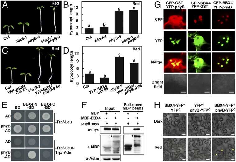
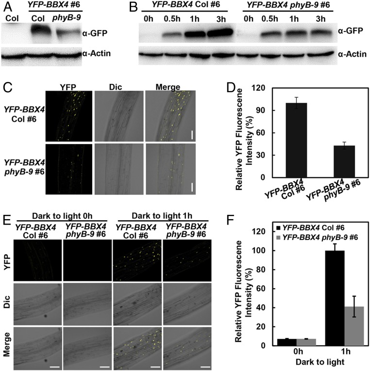
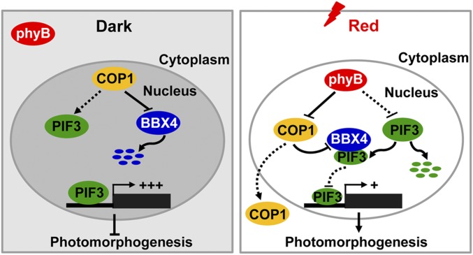
Phytochrome B (phyB) absorbs red light signals and subsequently initiates a set of molecular events in plant cells to promote photomorphogenesis. Here we show that phyB directly interacts with B-BOX CONTAINING PROTEIN 4 (BBX4), a positive regulator of red light signaling, and positively controls its abundance in red light. BBX4 associates with PHYTOCHROME INTERACTING FACTOR 3 (PIF3) and represses PIF3 transcriptional activation activity and PIF3-controlled gene expression. The degradation of BBX4 in darkness is dependent on CONSTITUTIVELY PHOTOMORPHOGENIC 1 (COP1) and the 26S proteasome system. Collectively, BBX4 acts as a key component of the phyB-PIF3-mediated signaling module and fine tunes the red light action. phyB promotes the accumulation of BBX4, which in turn serves to repress PIF3 action through direct physical interaction to promote photomorphogenic development in red light.

Keywords: BBX4; COP1; light signaling; photomorphogenesis; phyB.


Conflict of interest statement

The authors declare no competing interest.

Figures

Fig. 1.
Fig. 1.
phyB genetically and physically interacts with BBX4. (A and B) Hypocotyl phenotype (A) and length (B) of 4-d-old Col, bbx4-1, phyB-9, and bbx4-1 phyB-9 seedlings grown in R (115.8 μmol/m2/s) light. The unit of hypocotyl length is millimeters. The experiments were performed 3 times with similar results. The graphs depict one of these experiments. Error bars represent SE (n ≥ 20). Letters above the bars indicate significant differences (P < 0.05), as determined by 1-way ANOVA with Tukey’s post hoc analysis. (C and D) Hypocotyl phenotype (C) and length (D) of 4-d-old Col, YFP-BBX4 #6, phyB-9, and YFP-BBX4 phyB-9 #6 seedlings grown in R light (115.8 μmol/m2/s). The unit of hypocotyl length is millimeters. The experiments were performed 3 times, with similar results. The graphs depict 1 of these experiments. Error bars represent SE (n ≥ 20). Letters above the bars indicate significant differences (P < 0.05), as determined by 1-way ANOVA with Tukey’s post hoc analysis. (E) Yeast two-hybrid interactions between the BBX4 and phyB. (F) Semi-in vivo pull-down assay of BBX4 with phyB. Total plant protein was extracted from 4-d-old phyB-myc transgenic seedlings grown in R light (115.8 μmol/m2/s). Equal amounts of MBP and MBP-BBX4 proteins were added to total plant protein extracts. The asterisk indicates MBP-BBX4. Actin served as a negative control. (G) BBX4 and phyB colocalize to the nuclear bodies in tobacco cells. CFP-BBX4 and YFP-phyB were transiently coexpressed in tobacco leaves. CFP-GST and YFP-GST served as negative controls. (Scale bars: 5 µm.) (H) BiFC assay showing the interaction of BBX4 with phyB in R light. BBX4 and phyB were fused to the N- and C-terminal fragments of YFP (YFPN and YFPC, respectively). Unfused YFPN and YFPC fragments served as negative controls. (Scale bars: 20 μm.)
Fig. 2.
Fig. 2.
phyB stabilizes BBX4 in R light. (A) YFP-BBX4 protein levels in YFP-BBX4 Col #6 and YFP-BBX4 phyB-9 #6 grown in R light (115.8 μmol/m2/s) for 4 d. Col served as a negative control. (B) Immunoblot analysis of YFP-BBX4 protein levels in YFP-BBX4 Col #6 and YFP-BBX4 phyB-9 #6 grown in the dark for 4 d and then transferred to R light (115.8 μmol/m2/s) for 0, 0.5, 1, and 3 h, as indicated. Actin served as a loading control. (C and D) Analysis of YFP fluorescence signals in hypocotyls of YFP-BBX4 Col #6 and YFP-BBX4 phyB-9 #6 seedlings grown in R light (115.8 μmol/m2/s) for 4 d. The corresponding fluorescence intensity was measured using ImageJ and was compared between the overall signals from the images, as shown in D. Data are mean ± SE (n ≥ 10). (Scale bars: 100 μm.) (E and F) Analysis of YFP fluorescence signals in hypocotyls of YFP-BBX4 Col #6 and YFP-BBX4 phyB-9 #6 seedlings grown in the dark for 4 d, then transferred to R light (115.8 μmol/m2/s) for 0 and 1 h. The corresponding fluorescence intensity was measured using ImageJ software and compared between the overall signals from the images, as shown in F. Data are mean ± SE (n ≥ 10). (Scale bars: 100 μm.)
Fig. 3.
Fig. 3.
BBX4 genetically and physically interacts with PIF3. (A and B) Hypocotyl phenotype (A) and length (B) of 4-d-old Col, bbx4-1, pif3-1 and bbx4-1 pif3-1 seedlings grown in R light (115.8 μmol/m2/s). The unit of hypocotyl length is millimeters. The experiments were performed 3 times, with similar results. The graphs depict 1 of these experiments. Error bars represent SE (n ≥ 20). Letters above the bars indicate significant differences (P < 0.05), as determined by 1-way ANOVA with Tukey’s post hoc analysis. (C) Yeast two-hybrid interactions between the BBX4 and PIF3. (D) FRET between CFP-PIF3 and YFP-BBX4 analyzed by acceptor bleaching in nuclei. (Top) Representative prebleach nuclei coexpressing YFP-BBX4 and CFP-PIF3 excited with a 514-nm or 405-nm laser, resulting in emission from YFP or CFP, respectively. (Bottom) The same nuclei after bleaching excited with a 514-nm or 405-nm laser. (E) The relative intensities of both YFP and CFP inside the nuclei were measured once before and twice after the bleaching, as indicated in D. (F) BiFC assay showing the interaction of BBX4 with PIF3 in red light. BBX4 and PIF3 were fused to the N- and C-terminal fragments of YFP (YFPN and YFPC, respectively). Unfused YFPN and YFPC fragments served as negative controls. (Scale bars: 40 μm.)
Fig. 4.
Fig. 4.
BBX4 inhibits the transcriptional activation activity of PIF3. (A) Yeast one-hybrid analysis showing that BBX4 inhibits the ability of PIF3 to bind to BBX23 promoter. Error bars represent SD (n =3). **P < 0.01, Student’s t test. (B) Schematic representation of constructs used in the transient transfection assay in Arabidopsis protoplasts. Arrows after the 35S promoters indicate that the transcriptional start site BBX29 promoter was fused to the firefly luciferase to create the reporter construct. (C) Transient dual LUC reporter gene assay showing that BBX4 represses the transcriptional activity of PIF3 on proBBX29:LUC reporter. Error bars represent SD (n = 3). **P < 0.01, Student’s t test. (D) Expression levels of BBX23, BBX29, XTR7, SNRK2.5, SDR, and ARF18 in Col, pif3-1, bbx4-1, and bbx4-1pif3-1 mutants. All seedlings were grown in darkness for 3 d and then transferred to R light for 30 min. Error bars represent SD (n = 3).
Fig. 5.
Fig. 5.
A proposed working model depicting the mechanism of BBX4 in phyB-PIF3–mediated light signaling. In darkness, phyB is in inactive form in the cytoplasm. COP1 stabilizes PIF3 and interacts with BBX4 to promote its degradation via the 26S proteasome system. Highly accumulated PIF3 induces the expression of its direct-target genes to repress photomorphogenesis. On R light illumination, phyB is converted to a biologically active form and translocated into the nucleus. Photoactivated phyB promotes PIF3 protein degradation and induces the accumulation of BBX4 protein, likely by inhibiting the COP1–BBX4 association. In addition, accumulated BBX4 interacts with the remaining PIF3 to inhibit the transcription of PIF3 direct-target genes and promote photomorphogenesis.

References

    1. Sharrock R. A., Quail P. H., Novel phytochrome sequences in Arabidopsis thaliana: Structure, evolution, and differential expression of a plant regulatory photoreceptor family. Genes Dev. 3, 1745–1757 (1989). - PubMed
    1. Gallagher S., Short T. W., Ray P. M., Pratt L. H., Briggs W. R., Light-mediated changes in two proteins found associated with plasma membrane fractions from pea stem sections. Proc. Natl. Acad. Sci. U.S.A. 85, 8003–8007 (1988). - PMC - PubMed
    1. Lin C., et al. , Association of flavin adenine dinucleotide with the Arabidopsis blue light receptor CRY1. Science 269, 968–970 (1995). - PubMed
    1. Guo H., Yang H., Mockler T. C., Lin C., Regulation of flowering time by Arabidopsis photoreceptors. Science 279, 1360–1363 (1998). - PubMed
    1. Rizzini L., et al. , Perception of UV-B by the Arabidopsis UVR8 protein. Science 332, 103–106 (2011). - PubMed

Publication types

MeSH terms

Substances

LinkOut - more resources