. 2020 Jan 31;94(4):e01925-19.
doi: 10.1128/JVI.01925-19. Print 2020 Jan 31.

Nucleocapsid Protein Recruitment to Replication-Transcription Complexes Plays a Crucial Role in Coronaviral Life Cycle

Affiliations

Nucleocapsid Protein Recruitment to Replication-Transcription Complexes Plays a Crucial Role in Coronaviral Life Cycle

Yingying Cong et al. J Virol. .

Abstract

Coronavirus (CoV) nucleocapsid (N) proteins are key for incorporating genomic RNA into progeny viral particles. In infected cells, N proteins are present at the replication-transcription complexes (RTCs), the sites of CoV RNA synthesis. It has been shown that N proteins are important for viral replication and that the one of mouse hepatitis virus (MHV), a commonly used model CoV, interacts with nonstructural protein 3 (nsp3), a component of the RTCs. These two aspects of the CoV life cycle, however, have not been linked. We found that the MHV N protein binds exclusively to nsp3 and not other RTC components by using a systematic yeast two-hybrid approach, and we identified two distinct regions in the N protein that redundantly mediate this interaction. A selective N protein variant carrying point mutations in these two regions fails to bind nsp3 in vitro, resulting in inhibition of its recruitment to RTCs in vivo Furthermore, in contrast to the wild-type N protein, this N protein variant impairs the stimulation of genomic RNA and viral mRNA transcription in vivo and in vitro, which in turn leads to impairment of MHV replication and progeny production. Altogether, our results show that N protein recruitment to RTCs, via binding to nsp3, is an essential step in the CoV life cycle because it is critical for optimal viral RNA synthesis.IMPORTANCE CoVs have long been regarded as relatively harmless pathogens for humans. Severe respiratory tract infection outbreaks caused by severe acute respiratory syndrome CoV and Middle East respiratory syndrome CoV, however, have caused high pathogenicity and mortality rates in humans. These outbreaks highlighted the relevance of being able to control CoV infections. We used a model CoV, MHV, to investigate the importance of the recruitment of N protein, a central component of CoV virions, to intracellular platforms where CoVs replicate, transcribe, and translate their genomes. By identifying the principal binding partner at these intracellular platforms and generating a specific mutant, we found that N protein recruitment to these locations is crucial for promoting viral RNA synthesis. Moreover, blocking this recruitment strongly inhibits viral infection. Thus, our results explain an important aspect of the CoV life cycle and reveal an interaction of viral proteins that could be targeted in antiviral therapies.

Keywords: coronavirus; nucleocapsid N protein; replication-transcription complexes; viral mRNA synthesis.


Figures

FIG 1
FIG 1
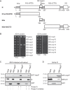
MHV N protein directly binds to nsp3. (A) Schematic structural organization of MHV N protein and overview of the truncations generated in this study. (B) The Y2H assay was used for analysis of the interactions between the N protein and each one of the MHV nsps. The plasmid expressing the AD-N fusion protein was cotransformed into the Y2H test strain with the plasmids carrying the nsp genes N-terminally tagged with the BD. Transformed cells were selected on a selective medium containing histidine (+His), while the interaction between the two tested proteins was assessed on a selective medium lacking histidine (−His) (42). Growth on plates without histidine showed that the N protein interacted only with nsp3. (C) Cell extracts from MHV-infected LR7 cells were incubated with RNase A or RNase A mixed with an RNase A inhibitor or were left untreated on ice for 30 min before being subjected to pulldown with immobilized GST or GST-nsp3N. Bound proteins were eluted by boiling in sample buffer and were analyzed by Western blotting using an anti-N antibody. Staining of the membrane with Ponceau red was used to reveal the amounts of immobilized GST and GST-nsp3N. (D) Bacterial lysates from E. coli cells expressing the 6×His-N fusion protein were prepared as described in Materials and Methods and were incubated with immobilized GST or GST-nsp3N. Bound proteins were eluted by boiling in sample buffer and were examined by Western blotting using an anti-His antibody. Amounts of immobilized GST and GST-nsp3N were visualized as in panel C.
FIG 2
FIG 2
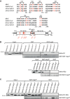
Two regions in the MHV N protein are required for its interaction with nsp3. (A) The interactions between nsp3N and different N protein forms with C-terminal truncations were tested with the Y2H assay, as described for Fig. 1B. (B) Bacterial extracts from E. coli cells expressing the 6×His-tagged N, N1, N2a, or N2b-N3 forms with truncations were incubated with immobilized GST or GST-nsp3N. Isolated proteins were eluted by boiling in sample buffer and were analyzed by Western blotting using an anti-6×His monoclonal antibody. GST and GST-nsp3N were visualized as in Fig. 1C. The asterisk indicates a degradation product of 6×His-N. (C) A bacterial extract from E. coli cells expressing the 6×His-tagged N2a was incubated with immobilized GST or GST-nsp3N. Isolated proteins were eluted and examined as in panel B, but Western blot membrane exposure times to visualize the bands were increased 5-fold.
FIG 3
FIG 3
Two distinct amino acid stretches mediate N protein interaction with nsp3. (A) (Top) The alignment of part of Betacoronavirus N protein sequences was performed using Jalview software (http://www.jalview.org/download.html). Conserved amino acids were localized on the three-dimensional structure of the N1-N2a domain (24) using PyMOL software (Schrödinger, New York, NY) to determine which ones are on the surface and thus available for interaction. The amino acids changed in the different mutant variants are indicated with an asterisk and colored in red. The Ser205 of MHV N protein, which represents the priming site essential for SR-rich region phosphorylation by host cell GSK-3, is colored in violet (32, 46). (Bottom) Schematic distribution of the different substitutions introduced in the N protein. (B) Bacterial extracts from E. coli cells expressing 6×His-tagged N1, N1m3, N1m4, N1m5, N1m6, N2a, N2am1, or N2am2 were incubated with immobilized GST or GST-nsp3N. Isolated proteins were eluted by boiling in sample buffer and were analyzed by Western blotting using an anti-6×His monoclonal antibody. GST and GST-nsp3N were visualized as in Fig. 1C. (C) Bacterial extracts from E. coli cells expressing 6×His-tagged N1-N2a, N1-N2am1, N1-N2am2, N1-N2am4, or N1-N2am6 were incubated with immobilized GST or GST-nsp3N. Isolated proteins were analyzed as in panel B.
FIG 4
FIG 4
In vitro binding defects of the doubly mutated N protein variants. (A) (Top). Schematic overview of the generated doubly mutated N protein variants. (Bottom) Bacterial extracts from E. coli cells expressing 6×His-tagged N, Nm4m1, Nm6m1, Nm4m2, or Nm6m2 were incubated with immobilized GST or GST-nsp3N. Isolated proteins were eluted by boiling in sample buffer and analyzed by Western blotting using an anti-6×His monoclonal antibody. GST and GST-nsp3N were visualized as in Fig. 1C. (B) Bacterial extracts from E. coli cells expressing 6×His-tagged Nm4m1 or Nm6m1 were incubated with immobilized GST or GST-N. Isolated proteins were examined as in panel A, and GST and GST-N protein were visualized as in Fig. 1C. (C) Bacterial extracts from E. coli cells expressing 6×His-tagged MHV N, Nm4m1, or Nm6m1 proteins were sedimented on a glycerol gradient of 15% to 40% at 135,000 × g for 75 min. Eleven fractions were collected from the top of the gel, and N protein chimera distribution over the gradient was analyzed using antibodies against the 6×His tag. (D) Quantification of the immunoblots from three independent fractionation experiments performed as in panel C. Error bars represent standard deviations.
FIG 5
FIG 5
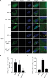
N protein localization to RTCs depends on its binding to nsp3. HeLa-CEACAM1a cells were transfected with plasmids expressing GFP, GFP-N, or GFP-Nm6m1 for 48 h before being infected or not with MHV for 7 h and processed for immunofluorescence using antibodies against MHV nsp2/nsp3. (A) Representative images of the experiment, which were quantified in panels B and C. DAPI, 4ʹ,6-diamidino-2-phenylindole. (B) The percentage of GFP-positive cells that were infected by MHV was calculated by counting the cells expressing nsp2/nsp3 and dividing that number by the number of GFP-positive cells. (C) The percentage of GFP-positive nsp2/nsp3 puncta was quantified in GFP-positive cells. Scale bar, 10 μm. Error bars represent standard deviations of three independent experiments. *, P < 0.05.
FIG 6
FIG 6
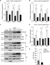
Host cells expressing the Nm6m1 variant have a dominant negative effect on MHV infection. (A) HeLa-CEACAM1a cells were transfected with plasmids expressing GFP, GFP-N, or GFP-Nm6m1 for 48 h and subsequently were infected with the MHV-2aFLS strain, at an MOI of 2.5, 5, or 10, for 7 h. In all of the experiments, 80% of the cells were transfected, as verified with a fluorescence microscope. Data represent the average luciferase activity, relative to HeLa-CEACAM1a cells expressing GFP, at each MOI. (B) HeLa-CEACAM1a cells were transfected as in panel A and subsequently were infected with the MHV-2aFLS strain, at an MOI of 5, for 5, 7, or 9 h. Data represent the average luciferase activity, relative to HeLa-CEACAM1a cells expressing GFP, at each infection time point. (C and D) HeLa-CEACAM1a cells, transfected as in panel A, were infected with MHV, at an MOI of 5, for 5, 7, or 9 h. Nontransfected cells not exposed to the virus were used as an extra control (mock). Proteins separated by SDS-PAGE were probed by Western blotting using antibodies recognizing MHV N protein, GFP, and actin (C), and viral N protein expression was quantified and normalized to that of actin (D). Data depicted in panel D represent N protein amounts, relative to HeLa-CEACAM1a cells expressing GFP, at each infection time point. (E) Production of virus progeny in experiments performed as in panel C. Cell culture supernatants were collected at 7 h p.i., and infectious virus progeny was assessed by determining the PFU per microliter of supernatant. Data represent virus titers, relative to HeLa-CEACAM1a cells expressing GFP, at each infection time point. Error bars represent standard errors of three (D and E) or four (A and B) independent experiments. *, P < 0.05.
FIG 7
FIG 7
The N protein-nsp3 interaction induces RTC-mediated viral RNA synthesis. (A) HeLa-CEACAM1a cells were transfected as in Fig. 6A and subsequently infected with the MHV-A59 strain, at an MOI of 5, for 7 h before quantification of the amount of gRNA and sgmRNA by RT-PCR, using the primer sets described in Table 1. The expression levels of gRNA and sgmRNA were normalized to those of GAPDH, and data represent average amounts, relative to HeLa-CEACAM1a cells expressing GFP. (B) Cleared cell lysates from LR7 cells infected with MHV for 7 h (Ext) were centrifuged at 13,000 × g for 10 min to obtain pellet (P13) and supernatant (S13) fractions. Equivalent amounts of each fraction were separated by SDS-PAGE and analyzed by Western blotting using antibodies against MHV N protein, VAP-A (Santa Cruz Biotechnology, Dallas, TX), and GAPDH (Fitzgerald Industries International, Acton, MA). (C) IVRS assays were performed with P13 fractions from mock-infected (P13) or MHV-infected (P13-MHV) cells, mixed with either dilution buffer or S13 from MHV-infected cells (S13-MHV). Synthesis of viral RNA was assessed by determining viral RNA levels by RT-PCR. Data are presented relative to those of the reaction with P13-MHV and dilution buffer. (D) The IVRS reactions were carried out with P13-MHV and S13-MHV, in the presence of 0, 0.1, 1, 2, or 5 μg of bacterial lysates from E. coli cells expressing 6×His-N. gRNA levels were measured and data are presented relative to the IVRS reaction in the absence of 6×His-N. (E) The IVRS assays were carried out with P13-MHV-infected cells, in the presence of 1 μg of bacterial lysates from E. coli cells expressing similar levels of 6×His-N or 6×His-Nm6m1, as verified prior to each experiment by Western blotting using an anti-6×His tag antibody. The viral RNA levels were measured and are presented relative to the control in which the IVRS reaction was performed in the absence of 6×His-N or 6×His-Nm6m1. Error bars represent standard deviations of three (A, C, and D) or four (E) independent experiments. *, P < 0.05.
FIG 8
FIG 8
The role of CoV N or Nm6m1 proteins at the viral replication platforms. Upon synthesis, CoV N or Nm6m1 proteins constitutively assemble into cytoplasmic oligomers. The wild-type N oligomers are recruited, via the interaction with nsp3, to the RTCs that are localized on DMVs and convoluted membranes. There, the N oligomers stimulate gRNA and sgmRNA synthesis. It may also be that the presence of N oligomers at the RTCs promotes local formation of ribonucleoprotein complexes. In contrast, the inability of Nm6m1 protein to be recruited to the RTCs severely impairs transcription, replication, and ultimately virion production.

References

    1. Cui J, Li F, Shi ZL. 2019. Origin and evolution of pathogenic coronaviruses. Nat Rev Microbiol 17:181–192. doi:10.1038/s41579-018-0118-9. - DOI - PMC - PubMed
    1. Perlman S, Netland J. 2009. Coronaviruses post-SARS: update on replication and pathogenesis. Nat Rev Microbiol 7:439–450. doi:10.1038/nrmicro2147. - DOI - PMC - PubMed
    1. Fehr AR, Perlman S. 2015. Coronaviruses: an overview of their replication and pathogenesis. Methods Mol Biol 1282:1–23. doi:10.1007/978-1-4939-2438-7_1. - DOI - PMC - PubMed
    1. Woo PC, Lau SK, Lam CS, Lau CC, Tsang AK, Lau JH, Bai R, Teng JL, Tsang CC, Wang M, Zheng BJ, Chan KH, Yuen KY. 2012. Discovery of seven novel mammalian and avian coronaviruses in the genus Deltacoronavirus supports bat coronaviruses as the gene source of Alphacoronavirus and Betacoronavirus and avian coronaviruses as the gene source of Gammacoronavirus and Deltacoronavirus. J Virol 86:3995–4008. doi:10.1128/JVI.06540-11. - DOI - PMC - PubMed
    1. Sawicki SG, Sawicki DL, Siddell SG. 2007. A contemporary view of coronavirus transcription. J Virol 81:20–29. doi:10.1128/JVI.01358-06. - DOI - PMC - PubMed

Publication types

Substances

LinkOut - more resources