

CircDUSP16 promotes the tumorigenesis and invasion of gastric cancer by sponging miR-145-5p
- PMID: 31776711
- PMCID: PMC7165161
- DOI: 10.1007/s10120-019-01018-7
CircDUSP16 promotes the tumorigenesis and invasion of gastric cancer by sponging miR-145-5p
Abstract
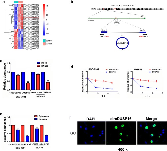
Background: Circular RNAs (circRNAs) as a novel subgroup of non-coding RNAs act a critical role in the pathogenesis of gastric cancer (GC). However, the underlying mechanisms by which hsa_circ_0003855 (circDUSP16) contributes to GC are still undocumented.
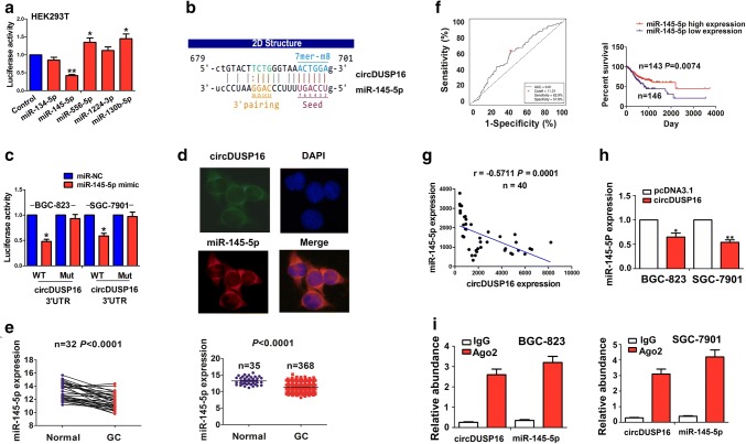
Materials: The differentially expressed circRNAs were identified by GEO database. The association of circDUSP16 or miR-145-5p expression with clinicopathological features and prognosis in GC patients was analyzed by FISH and TCGA-seq data set. Loss- and gain-of-function experiments as well as a xenograft tumor model were performed to assess the role of circDUSP16 in GC cells. circDUSP16-specific binding with miR-145-5p was confirmed by bioinformatic analysis, luciferase reporter, and RNA immunoprecipitation assays.
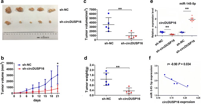
Results: The expression levels of circDUSP16 were markedly increased in GC tissue samples and acted as an independent prognostic factor of poor survival in patients with GC. Knockdown of circDUSP16 repressed the cell viability, colony formation, and invasive potential in vitro and in vivo, but ectopic expression of circDUSP16 reversed these effects. Moreover, circDUSP16 possessed a co-localization with miR-145-5p in the cytoplasm, and acted as a sponge of miR-145-5p, which attenuated circDUSP16-induced tumor-promoting effects and IVNS1ABP expression in GC cells. MiR-145-5p had a negative correlation with circDUSP16 expression and its low expression was associated with poor survival in GC patients.
Conclusions: CircDUSP16 facilitates the tumorigenesis and invasion of GC cells by sponging miR-145-5p, and may provide a novel therapeutic target for GC.
Keywords: Gastric cancer; Growth; Invasion; circDUSP16; miR-145-5p.
Conflict of interest statement
The authors declare that they have no conflict of interest.
Figures


Similar articles
-
CircDUSP16 Contributes to Cell Development in Esophageal Squamous Cell Carcinoma by Regulating miR-497-5p/TKTL1 Axis.J Surg Res. 2021 Apr;260:64-75. doi: 10.1016/j.jss.2020.11.052. Epub 2020 Dec 14. J Surg Res. 2021. PMID: 33326930
-
Circular RNA hsa_circ_0004872 inhibits gastric cancer progression via the miR-224/Smad4/ADAR1 successive regulatory circuit.Mol Cancer. 2020 Nov 10;19(1):157. doi: 10.1186/s12943-020-01268-5. Mol Cancer. 2020. PMID: 33172486 Free PMC article.
-
Circular RNA hsa_circ_0001829 promotes gastric cancer progression through miR-155-5p/SMAD2 axis.J Exp Clin Cancer Res. 2020 Dec 11;39(1):280. doi: 10.1186/s13046-020-01790-w. J Exp Clin Cancer Res. 2020. Retraction in: J Exp Clin Cancer Res. 2023 Apr 26;42(1):101. doi: 10.1186/s13046-023-02673-6. PMID: 33308284 Free PMC article. Retracted.
-
CircRNAs in gastric cancer: current research and potential clinical implications.FEBS Lett. 2021 Nov;595(21):2644-2654. doi: 10.1002/1873-3468.14196. Epub 2021 Oct 21. FEBS Lett. 2021. PMID: 34561854 Review.
-
Contribution of circRNAs in gastric cancer.Pathol Res Pract. 2021 Nov;227:153640. doi: 10.1016/j.prp.2021.153640. Epub 2021 Sep 28. Pathol Res Pract. 2021. PMID: 34624593 Review.
Cited by
-
Bupivacaine suppresses the progression of gastric cancer through regulating circ_0000376/miR-145-5p axis.BMC Anesthesiol. 2020 Oct 30;20(1):275. doi: 10.1186/s12871-020-01179-4. BMC Anesthesiol. 2020. PMID: 33126850 Free PMC article.
-
Promoter Hypomethylation and miR-145-5p Downregulation- Mediated HDAC11 Overexpression Promotes Sorafenib Resistance and Metastasis of Hepatocellular Carcinoma Cells.Front Cell Dev Biol. 2020 Aug 12;8:724. doi: 10.3389/fcell.2020.00724. eCollection 2020. Front Cell Dev Biol. 2020. PMID: 32903337 Free PMC article.
-
Molecular Insight into Gastric Cancer Invasion-Current Status and Future Directions.Cancers (Basel). 2023 Dec 21;16(1):54. doi: 10.3390/cancers16010054. Cancers (Basel). 2023. PMID: 38201481 Free PMC article. Review.
-
Identification of a novel circRNA, hsa_circ_0065898, that regulates tumor growth in cervical squamous cell carcinoma.Transl Cancer Res. 2021 Jan;10(1):47-56. doi: 10.21037/tcr-20-2808. Transl Cancer Res. 2021. PMID: 35116238 Free PMC article.
-
MiR-145-5p overexpression rejuvenates aged adipose stem cells and accelerates wound healing.Biol Open. 2024 Feb 15;13(2):bio060117. doi: 10.1242/bio.060117. Epub 2024 Feb 19. Biol Open. 2024. PMID: 38315073 Free PMC article.
References
-
- Figueiredo C, Costa S, Karameris A, Machado JC. Pathogenesis of Gastric Cancer. Helicobacter. Suppl. 2015;1:30–35. - PubMed
MeSH terms
Substances
Grants and funding
LinkOut - more resources
Full Text Sources
Medical
Research Materials
Miscellaneous
