. 2019 May:25:10-20.
doi: 10.1016/j.mattod.2019.02.015. Epub 2019 Mar 9.

All-optical reconfigurable chiral meta-molecules

Affiliations

All-optical reconfigurable chiral meta-molecules

Linhan Lin et al. Mater Today (Kidlington). 2019 May.

Abstract

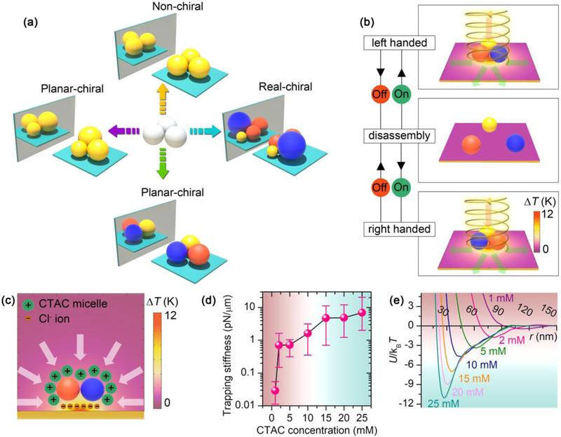
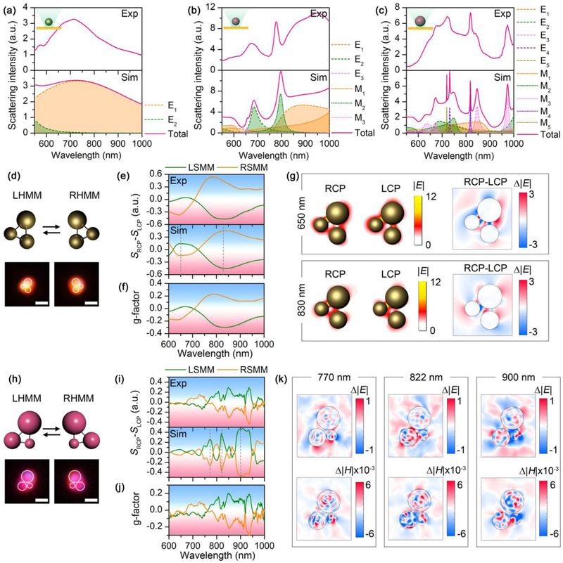
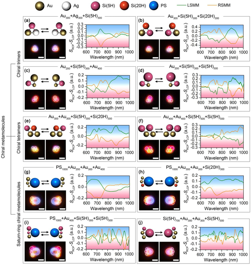
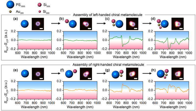
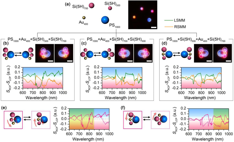
Chirality is a ubiquitous phenomenon in the natural world. Many biomolecules without inversion symmetry such as amino acids and sugars are chiral molecules. Measuring and controlling molecular chirality at a high precision down to the atomic scale are highly desired in physics, chemistry, biology, and medicine, however, have remained challenging. Herein, we achieve all-optical reconfigurable chiral meta-molecules experimentally using metallic and dielectric colloidal particles as artificial atoms or building blocks to serve at least two purposes. One is that the on-demand meta-molecules with strongly enhanced optical chirality are well-suited as substrates for surface-enhanced chiroptical spectroscopy of chiral molecules and as active components in optofluidic and nanophotonic devices. The other is that the bottom-up-assembled colloidal meta-molecules provide microscopic models to better understand the origin of chirality in the actual atomic and molecular systems.

Keywords: bottom-up assembly; metamolecules; optical chirality; opto-thermoelectric tweezers.


Figures

FIGURE 1.
FIGURE 1.
Concept and working principle of reconfigurable chiral meta-molecules. (a) Chirality origin in colloidal meta-molecules. The balls with different colors represent colloids with different materials. (b) Working principle of the reconfigurable opto-thermophoretic assembly of chiral meta-molecules. The red and green arrows represent the incident and scattering light, respectively. (c) Colloid-micelle interaction in the light-generated temperature field. (d) Measured trapping stiffness of single 300 nm AuNSs measured at different CTAC concentrations. (e) Interparticle interaction potential U between two 300 nm AuNSs at different CTAC concentrations. The green color indicates stable trapping or assembly region, while red color indicates that the trapping or assembly is not stable. T is the temperature and kB is the Boltzmann constant.
FIGURE 2.
FIGURE 2.
Building blocks of the chiral meta-molecules, and the Au and Si chiral trimers. (a-c) Experimental (top) and simulated (bottom) scattering spectra of single 260 nm AuNP (a), 300 nm SiNP (5H) (b), and 500 nm SiNP (5H) (c). Multipole decomposition is carried out for the simulated spectra, with the electrical (El and magnetic (Ml spherical Mie modes of l orders, i.e., l=1: dipole mode; l=2: quadrupole mode; l=3: octupole mode; l=4: hexadecapole mode, etc. (d) Schematic and optical images of left-handed and right-handed Au chiral trimer. (e) Experimental (top) and simulated (bottom) differential scattering spectra of the Au chiral trimer. (f) G-factor of the Au chiral trimer. (g) Electric field distribution and differential electric field distribution of the Au chiral trimer at the wavelengths of 650 nm (top) and 830 nm (bottom). (h) Schematic and optical images of left-handed and right-handed Si chiral trimer. (i) Experimental (top) and simulated (bottom) differential scattering spectra of the Si chiral trimer. (j) G-factor of the Si chiral trimer. (k) Differential electric and magnetic field distribution profiles of the Si chiral trimer at 770 nm (left), 822 nm (middle) and 900 nm (right). In (d-g), the diameters of the AuNPs are 230 nm, 260 nm, and 330 nm, respectively. In (h-k), the diameters of the SiNPs are 300 nm, 500 nm, and 700 nm, respectively. In (d) and (h), white circles are superimposed onto the dark-field optical images to guide the visualization of the molecular structures. Scale bars: 1 μm.
FIGURE 3.
FIGURE 3.
Versatility of the experimentally assembled chiral meta-molecules. Schematic, optical images and differential scattering spectra of chiral trimers composed of (a) an AuNP, an AgNP and a SiNP (5H) with diameter of 300 nm; (b) an AuNP, a SiNP (5H) and a SiNP (20H) with diameter of 300 nm; (c) a 200 nm AuNP, a 300 nm SiNP (5H), and a 400 nm AuNP; (d) a 200 nm AuNP, a 300 nm SiNP (5H) and a 500 nm SiNP (5H). Schematic, optical images and differential scattering spectra of chiral tetramers composed of (e) two 300 nm AuNP, a 300 nm SiNP (5H), and a 300 nm SiNP (20H); (f) two 400 nm AuNPs and two 500 nm SiNPs (5H). Schematic, optical images and differential scattering spectra of Saturn-ring chiral meta-molecules composed of (g) a 1 μm PS bead, a 200 nm AuNP, a 300 nm AuNP, and a 400 nm AuNP; (h) a 1 μm PS bead, two 300 nm AuNPs, and a 300 nm SiNP (20H); (i) a 1 μm PS bead, a 400 nm AuNP, a 300 nm SiNP (5H), and a 500 nm SiNP (5H); (j) a 700 nm SiNP (5H), two 300 nm AuNPs, and a 300 nm SiNP (5H). The meta-molecules on the left are LHMMs, while the ones on the right are RHMMs. White circles are superimposed onto the dark-field optical images to guide the visualization of the molecular structures. Scale bar: 1 μm.
FIGURE 4.
FIGURE 4.
Assembly process of chiral meta-molecules of both handedness with in-situ measurement of the optical chirality. The schematics, optical images, and differential scattering spectra show the step-by-step assembly process of both the left-handed (a-d) and right-handed (e-h) chiral meta-molecules composed of a 1 μm PS, a 300 nm AuNP, a 300 nm SiNP (5H) and a 500 nm SiNP (5H). White circles are superimposed onto the dark-field optical images to guide the visualization of the molecular structures. Scale bar: 1 μm.
FIGURE 5.
FIGURE 5.
Reconfigurability of the experimentally assembled chiral meta-molecules. (a) Schematic and optical images of the dispersed meta-atoms. (b-d) Schematic, optical images, and differential scattering spectra of three sets of chiral meta-molecules composed of a 1 μm PS bead, a 400 nm AuNP, a 500 nm SiNP (5H), and a 700 nm SiNP (5H) with different configurations. (e-f) Comparison of the differential scattering spectra among the chiral meta-molecules. The solid and dashed curves correspond to the structures with solid and dashed outlines, respectively. In (b-d), the meta-molecules on the left are LHMMs, while the ones on the right are RHMMs. In (b-d), white circles are superimposed onto the dark-field optical images to guide the visualization of the molecular structures. In (e) and (f), the region superimposed with yellow shows mirror symmetry in the differential scattering spectra. Scale bars: 2 μm in (a); 1 μm in (b-d).

References

    1. Wang Z, et al., Adv. Mater 29 (27) (2017) 1700412. - PubMed
    1. Hu T, et al., Nano Lett. 14 (12) (2014) 6799. - PubMed
    1. Yeom J, et al., Nat. Mater 14 (1) (2015) 66. - PMC - PubMed
    1. Ackerman PJ, and Smalyukh II, Nat. Mater 16 (2016) 426. - PubMed
    1. Ito TH, et al., Langmuir 32 (33) (2016) 8461. - PubMed

LinkOut - more resources