. 2019 Jun 6;1(4):267-276.
doi: 10.1002/acr2.1039. eCollection 2019 Jun.

The NLRP3 p.A441V Mutation in NLRP3-AID Pathogenesis: Functional Consequences, Phenotype-Genotype Correlations and Evidence for a Recurrent Mutational Event

Affiliations

The NLRP3 p.A441V Mutation in NLRP3-AID Pathogenesis: Functional Consequences, Phenotype-Genotype Correlations and Evidence for a Recurrent Mutational Event

Fawaz Awad et al. ACR Open Rheumatol. .

Abstract

Objective: To determine the molecular and cellular bases of autoinflammatory syndromes in a multigenerational French family with Muckle-Wells syndrome and in a patient originating from Portugal with familial cold autoinflammatory syndrome.

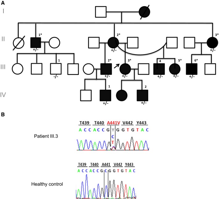
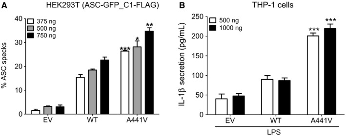
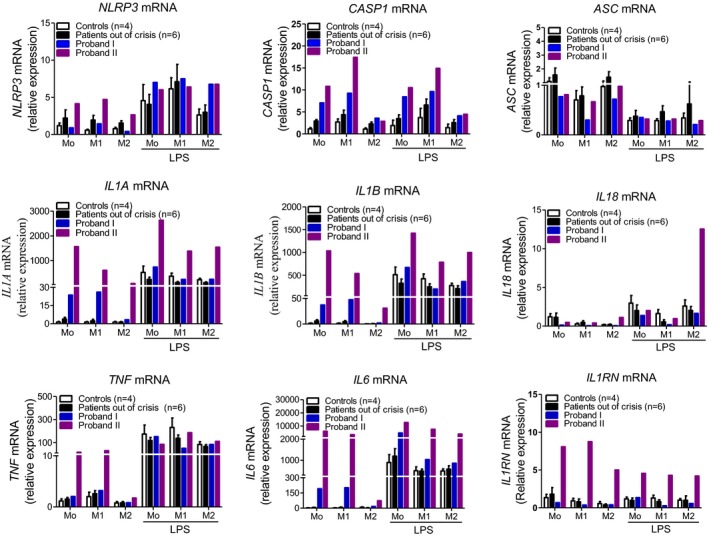
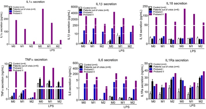
Methods: Sequencing of NLRP3 exon 3 was performed in all accessible patients. Microsatellite and whole-genome single nucleotide polymorphism genotyping was used i) to test the intrafamilial segregation of the identified variant and ii) to look for a founder effect. Functional analyses included the study of i) apoptosis-associated speck-like protein containing a CARD (ASC) speck formation in HEK293T cells (stably expressing ASC-green fluorescent protein and pro-caspase 1-FLAG) transiently expressing the wild-type or mutated NLRP3 protein, ii) levels of IL-1β secreted from transfected THP-1 cells, and iii) inflammasome-related gene expression and cytokine secretion from monocytes isolated from patients in crisis (probands from the two families), related patients out of crisis, and from controls.

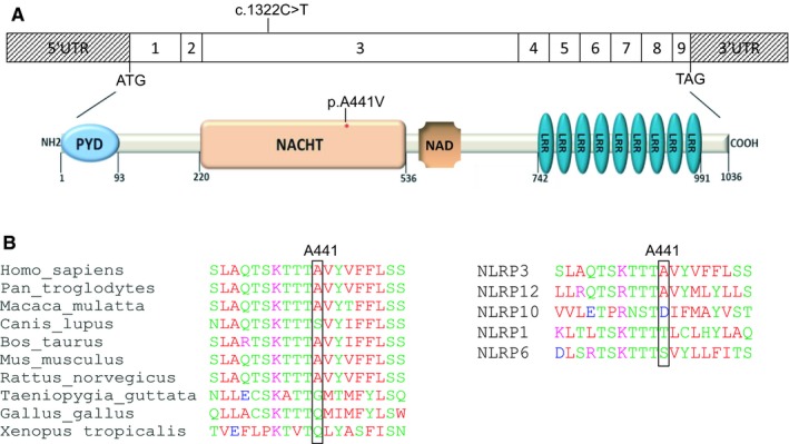
Results: The same heterozygous mutation (c.1322C>T, p.A441V) located in the NACHT domain, segregating with the disease within the first family, was identified in the two families. This mutation was found to be associated with different core haplotypes. NLRP3-A441V led to increased ASC speck formation and high levels of secreted IL-1β. Monocyte inflammasome-related gene expression and cytokine secretion, which were within the normal range in patients out of crisis, were found to be differentially regulated between the two probands, correlating with their phenotypic status.

Conclusion: These molecular and cellular findings, which indicate a recurrent mutational event, clearly demonstrate the pathogenicity of the p.A441V missense mutation in NLRP3-associated autoinflammatory disease and point to the interest of studying patients' primary cells to assess disease activity.

Keywords: IL‐1β; NLRP3; NLRP3‐AID; cryopyrin‐associated periodic syndromes; inflammasome; variation.


Figures

Figure 1
Figure 1
A, Genealogical tree of Family I with NLRP3AID and the identified NLRP3 mutation c.1322C>T (p.A441V). Filled symbols represent affected individuals, and empty symbols represent healthy subjects. The black arrow shows Proband I. The asterisk indicates that blood was available for monocyte isolation and ex vivo study. Nucleotide and amino acid coordinates are given assuming that the A of the first ATG codon is nucleotide #1. B, NLRP3 electropherograms corresponding to the direct Sanger sequencing of the polymerase chain reaction (PCR) products from one of the NLRP3AID patients (individual III.3, upper panel) showing the heterozygous mutation (c.1322C>T, p.A441V) and a control (lower panel).
Figure 2
Figure 2
A, Impact of the NLRP3 c.1322C>T transition at the protein level. Exonic organization of the human NLRP3 cDNA and domain‐organization model of the corresponding protein. The identified mutation is indicated. The red asterisk shows the location of the identified mutation in the NACHT domain (top). PYD, pyrin domain; NACHT (NAIP,CIITA,HET‐E and TP1) domain; NAD, NACHT‐associated domain; LRR, leucine‐rich repeat domain. B, Partial interspecies alignment of NLRP3 showing the evolutionary conservation of the domain containing A441 (left panel), as well as the conservation of A441 in NLRP12, one of the human NLRP3 paralogs (right panel).
Figure 3
Figure 3
A, Impact of the c.1322C>T, (p.A441V) mutation on apoptosis‐associated speck‐like protein containing a CARD (ASC) speck formation. HEK293T (ASCGFP_C1‐FLAG) cells were transfected with different amounts (375, 500, or 750 ng) of the pNLRP3‐WT, pNLRP3‐A441V, or the empty vector (EV). The percentage of ASC specks was calculated as described in Materials and Methods. Results represent the means ± SD from three independent experiments. P values were calculated using unpaired student's t test. Asterisks indicates significant P value (<0.05) as compared with the wild‐type (***P value = 0.0009, *P value = 0.0113, **P value = 0.0031). B, IL‐1β secretion, as assessed by ELISA in cell culture supernatants of phorbol myristate acetate–primed THP‐1 cells transfected with either 500 or 1000 ng of pNLRP3‐WT, pNLRP3‐A441V, or the empty vector and treated with LPS, a known stimulus for NLRP3 inflammasome. Results represent the mean ± SD of 3 independent experiments performed in triplicates. P values were calculated using unpaired student's t test. Asterisks indicates significant P value (<0.05) as compared with the wild type (***P value = 0.0009 or P value = 0.0006).
Figure 4
Figure 4
Expression of inflammasome‐related genes (NLRP3, CASP1, and ASC) and genes encoding cytokines in human polarized monocytes (M1: proinflammatory phenotype, M2: anti‐inflammatory phenotype, M0: basal conditions) in the presence or absence of LPS in Proband I, patients out of crisis (n = 6), and Proband II, as compared with apparently healthy donors (n = 4).
Figure 5
Figure 5
Cytokine levels (IL‐1β, IL‐1α, IL‐18, TNF‐α, IL‐6 and IL‐1Ra) in the supernatant of polarized monocytes (M1: proinflammatory phenotype, M2: anti‐inflammatory phenotype, M0: basal conditions) of Proband I, patients out of crisis (n = 6), and Proband II as compared with apparently healthy donors (n = 4) in the presence or absence of LPS.

References

    1. Ben‐Chetrit E, Gattorno M, Gul A, Kastner DL, Lachmann HJ, Touitou I, et al. Consensus proposal for taxonomy and definition of the autoinflammatory diseases (AIDs): a Delphi study. Ann Rheum Dis 2018;77:1558–65. - PubMed
    1. Kuemmerle‐Deschner JB. CAPS–pathogenesis, presentation and treatment of an autoinflammatory disease. Semin Immunopathol 2015;37:377–85. - PubMed
    1. Hoffman HM, Mueller JL, Broide DH, Wanderer AA, Kolodner RD. Mutation of a new gene encoding a putative pyrin‐like protein causes familial cold autoinflammatory syndrome and Muckle‐Wells syndrome. Nat Genet 2001;29:301–5. - PMC - PubMed
    1. Feldmann J, Prieur AM, Quartier P, Berquin P, Certain S, Cortis E, et al. Chronic infantile neurological cutaneous and articular syndrome is caused by mutations in CIAS1, a gene highly expressed in polymorphonuclear cells and chondrocytes. Am J Hum Genet 2002;71:198–203. - PMC - PubMed
    1. Martinon F, Mayor A, Tschopp J. The inflammasomes: guardians of the body. Annu Rev Immunol 2009;27:229–65. - PubMed

LinkOut - more resources