

Mechanisms linking adipose tissue inflammation to cardiac hypertrophy and fibrosis
- PMID: 31777927
- PMCID: PMC7191542
- DOI: 10.1042/CS20190578
Mechanisms linking adipose tissue inflammation to cardiac hypertrophy and fibrosis
Abstract
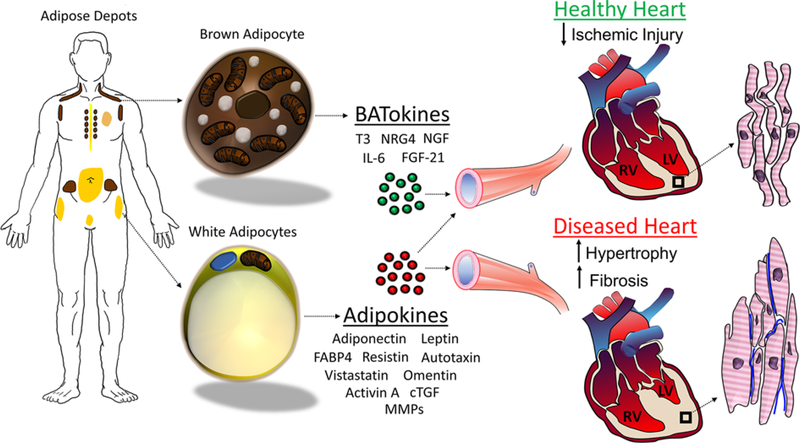
Adipose tissue is classically recognized as the primary site of lipid storage, but in recent years has garnered appreciation for its broad role as an endocrine organ comprising multiple cell types whose collective secretome, termed as adipokines, is highly interdependent on metabolic homeostasis and inflammatory state. Anatomical location (e.g. visceral, subcutaneous, epicardial etc) and cellular composition of adipose tissue (e.g. white, beige, and brown adipocytes, macrophages etc.) also plays a critical role in determining its response to metabolic state, the resulting secretome, and its potential impact on remote tissues. Compared with other tissues, the heart has an extremely high and constant demand for energy generation, of which most is derived from oxidation of fatty acids. Availability of this fatty acid fuel source is dependent on adipose tissue, but evidence is mounting that adipose tissue plays a much broader role in cardiovascular physiology. In this review, we discuss the impact of the brown, subcutaneous, and visceral white, perivascular (PVAT), and epicardial adipose tissue (EAT) secretome on the development and progression of cardiovascular disease (CVD), with a particular focus on cardiac hypertrophy and fibrosis.
Keywords: adipokines; brown adipose tissue; cardiac fibrosis; cardiac hypertrophy; cardiovascular disease; obesity.
© 2019 The Author(s). Published by Portland Press Limited on behalf of the Biochemical Society.
Conflict of interest statement
Competing Interests
The authors declare that there are no competing interests associated with the manuscript.
Figures
References
Publication types
MeSH terms
Grants and funding
LinkOut - more resources
Full Text Sources