Review
. 2020 Nov 24;44(6):655-683.
doi: 10.1093/femsre/fuz028.

Structure and genetics of Escherichia coli O antigens

Affiliations
Review

Structure and genetics of Escherichia coli O antigens

Bin Liu et al. FEMS Microbiol Rev. .

Abstract

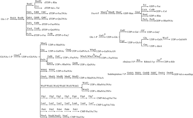
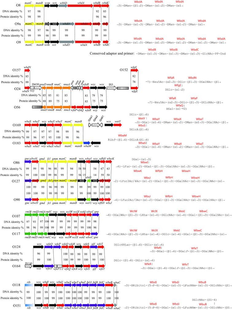
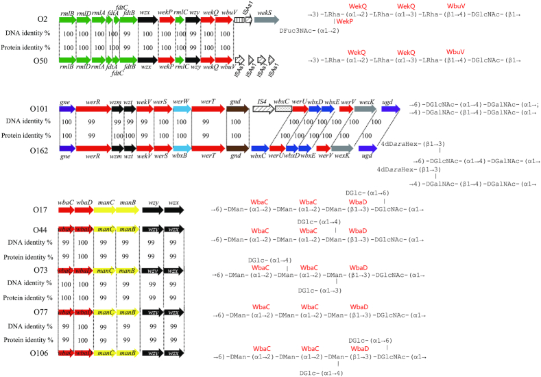
Escherichia coli includes clonal groups of both commensal and pathogenic strains, with some of the latter causing serious infectious diseases. O antigen variation is current standard in defining strains for taxonomy and epidemiology, providing the basis for many serotyping schemes for Gram-negative bacteria. This review covers the diversity in E. coli O antigen structures and gene clusters, and the genetic basis for the structural diversity. Of the 187 formally defined O antigens, six (O31, O47, O67, O72, O94 and O122) have since been removed and three (O34, O89 and O144) strains do not produce any O antigen. Therefore, structures are presented for 176 of the 181 E. coli O antigens, some of which include subgroups. Most (93%) of these O antigens are synthesized via the Wzx/Wzy pathway, 11 via the ABC transporter pathway, with O20, O57 and O60 still uncharacterized due to failure to find their O antigen gene clusters. Biosynthetic pathways are given for 38 of the 49 sugars found in E. coli O antigens, and several pairs or groups of the E. coli antigens that have related structures show close relationships of the O antigen gene clusters within clades, thereby highlighting the genetic basis of the evolution of diversity.

Keywords: Escherichia coli; O antigen; diversity; gene cluster; serogroup; structure.


Figures

Scheme 1.
Scheme 1.
Biosynthesis pathways for the sugars in E. coli O antigens. RmlA, glucose-1-phosphate thymidylyltransferase (Zuccotti et al  2001); RmlB, dTDP-d-glucose 4,6-dehydratase (Allard et al. 2001); RmlC, dTDP-4-keto-6-deoxy-d-glucose 3,5-epimerase (Giraud et al. 1999a); RmlD, dTDP-6-deoxy-l-mannose-dehydrogenase (Giraud et al. 1999b); ManA, phosphomannose isomerase; ManB, phosphomannomutase; ManC, mannose-1-phosphate guanylyltransferase (Samuel and Reeves 2003); Psb1, C6 dehydratase/C5 epimerase; Psb2, aminotransferase; Psb4, nucleotidase; Psb6, condensase; Psb3, cytidylyltransferase; FnlA, 4,6-dehydratase, 3- and 5-epimerase; FnlB, reductase; FnlC, C-2 epimerase (Kneidinger et al. 2003b); GalU, UTP-glucose-1-phosphate uridylyltransferase (Bonofiglio, Garcia and Mollerach 2005); GlmU, UDP-N-acetyl-glucosamine pyrophosphorylase (Mengin-Lecreulx and van Heijenoort 1993); GalE, UDP-glucose-4-epimerase (Samuel and Reeves 2003); Glf, UDP-galactopyranose mutase (Nassau et al. 1996); Gna, UDP-GalNAcA synthetase (Zhao et al. 2000); Gne, UDP-N-acetylglucosamine-4-epimerase (Bengoechea et al. 2002); Gae, C5-epimerase; Gla, UDP-galacturonatenase (Munoz et al. 1999); Ugd, UDP-glucose 6-dehydrogenase (Stevenson et al. 1996); QnlA, dTDP-4-dehydrorhamnose reductase; QnlB, C-2 epimerase (Kneidinger et al. 2003); Gmd, GDP-mannose-4,6-dehydratase (Somoza et al. ; Kneidinger et al. 2001); Fcl, GDP-l-fucose synthetase (Rosano et al. 2000); VioA, aminotransferase (Wang et al. 2007); VioB, N-acetyltransferase (Wang et al. 2007); ColA, GDP-4-keto-6-deoxy-d-mannose 3-dehydrase (Alam, Beyer and Liu 2004); ColB, GDP-colitose synthase (Alam, Beyer and Liu 2004); PerA, GDP-perosamine synthetase (Zhao et al. ; Albermann and Beuttler 2008); PerB, GDP-perosamine N-acetyltransferase (Albermann and Beuttler 2008); Rib, ribulose 5-phosphate reductase/CDP-ribitol pyrophosphorylase (Follens et al. 1999); FdtA, dTDP-6-deoxy-hex-4-ulose isomerase; FdtB, dTDP-6-deoxy-d-xylo-hex-3-ulose aminase; FdtC, dTDP-d-Fuc3N acetylase (Pfoestl et al. 2003); QdtA, dTDP-4-oxo-6-deoxy-d-glucose3,4-oxoisomerase; QdtB, dTDP-3-oxo-6-deoxy-d-glucose aminase (Pfostl et al. 2008); QdtC, dTDP-d-Qui3N acetylase (Pfostl et al. 2008); MnaA, UDP-N-acetylglucosamine-2-epimerase (Campbell et al. 2000); FcfA, C-4 reductase; FcfB, mutase; GmhA, Glyceromannoheptose-7-P isomerase; HddA, d-Heptose 7-phosphate kinase; GmhB, d-Heptose 1,7-biphosphate phosphatase; HddC, d-Heptose 1-phosphate guanosyltransferase; DmhA, NAD-dependentepimerase/dehydratase; DmhB, NAD-dependent epimerase/dehydratase; FdfA, aminotransferase; FdfB, acetyltransferase; Rmd, oxidoreductase; WbpS, aminotransferase; WbuX, aminotransferase; wbhS, UDP-d-GlcNAc 4,6-dehydratase; wbhP, NAD-dependent epimerase/dehydratase; GnaA, UDP-GlcNAc 6-dehydrogenase; MndA, oxidoreductase; MndB, acetyltransferase; MndC, aminotransferase; MndD, UDP-N-acetylglucosamine 2-epimerase; WeiP/WekE, aminotransferase; WeiQ/WekF, aminotransferase; WeiS/WekG, dehydratase/epimerase; WeiO, acetyltransferase; WekD, formyltransferase; NnaA, UDP-N-acetylglucosamine-2-epimerase; NnaB, N-acetylneuraminic acid synthetase; NnaC, CMP-N-acetylneuraminic acid synthetase; NnaD, N-acetylneuraminic acid synthetase; Elg1, UDP-N-acetylglucosamine 4,6-dehydratase; Elg2, aminotransferase; Elg3, UDP-N-acetylglucosamine 2-epimerase; Elg4, N-acetyl-neuraminic acid synthetase; Elg5, acetyltransferase; Elg6, sugar-phosphate nucleotide transferase, Elg7, cytidylyl-transferase; Lea1, C6 dehydratase; Lea2, aminotransferase; Lea3, alanyltransferase; Lea4, N-acetylneuraminic acid synthetase; Lea5, UDP-N-acetylglucosamine 2-epimerase; Lea6, sugar-phosphate nucleotide transferase; Lea7, CMP-N-acetylneuraminic acid synthetase; TarI, cytidylyltransferase. aThe enzyme is encoded by the gene which is not located in the O antigen gene cluster. bThe enzyme could not be assigned functionally.
Figure 1.
Figure 1.
O antigen gene clusters and structures that are identical or closely related within E. coli (A) KO5 and O8; KO3, O9 and O9a; O157, O24 and O56; O169 and O183; (B) O86, O127 and O90; O107 and O117; O124 and O164; O118 and O151; (C) O2 and O50; O101 and O162; O17, O44, O73, O77 and O106. For gene key, see Supplementary Fig. S1.
Figure 1.
Figure 1.
O antigen gene clusters and structures that are identical or closely related within E. coli (A) KO5 and O8; KO3, O9 and O9a; O157, O24 and O56; O169 and O183; (B) O86, O127 and O90; O107 and O117; O124 and O164; O118 and O151; (C) O2 and O50; O101 and O162; O17, O44, O73, O77 and O106. For gene key, see Supplementary Fig. S1.

References

    1. Achtman M, Pluschke G. Clonal analysis of descent and virulence among selected Escherichia coli. Annu Rev Microbiol. 1986;40:185–210. - PubMed
    1. Adeyeye A, Jansson P-E, Lindberg B et al. Structural studies of the Escherichia coli O-149 O-antigen polysaccharide. Carbohydr Res. 1988;176:231–6. - PubMed
    1. Al-Dabbagh B, Olatunji S, Crouvoisier M et al. Catalytic mechanism of MraY and WecA, two paralogues of the polyprenyl-phosphate N-acetylhexosamine 1-phosphate transferase superfamily. Biochimie. 2016;127:249–57. - PubMed
    1. Alam J, Beyer N, Liu HW. Biosynthesis of colitose: expression, purification, and mechanistic characterization of GDP-4-keto-6-deoxy-D-mannose-3-dehydrase (ColD) and GDP-L-colitose synthase (ColC). Biochemistry. 2004;43:16450–60. - PubMed
    1. Albermann C, Beuttler H. Identification of the GDP-N-acetyl-D-perosamine producing enzymes from Escherichia coli O157:H7. FEBS Lett. 2008;582:479–84. - PubMed

Publication types

MeSH terms