. 2020 Feb:324:113135.
doi: 10.1016/j.expneurol.2019.113135. Epub 2019 Nov 26.

The p53 inactivators pifithrin-μ and pifithrin-α mitigate TBI-induced neuronal damage through regulation of oxidative stress, neuroinflammation, autophagy and mitophagy

Affiliations

The p53 inactivators pifithrin-μ and pifithrin-α mitigate TBI-induced neuronal damage through regulation of oxidative stress, neuroinflammation, autophagy and mitophagy

Ling-Yu Yang et al. Exp Neurol. 2020 Feb.

Abstract

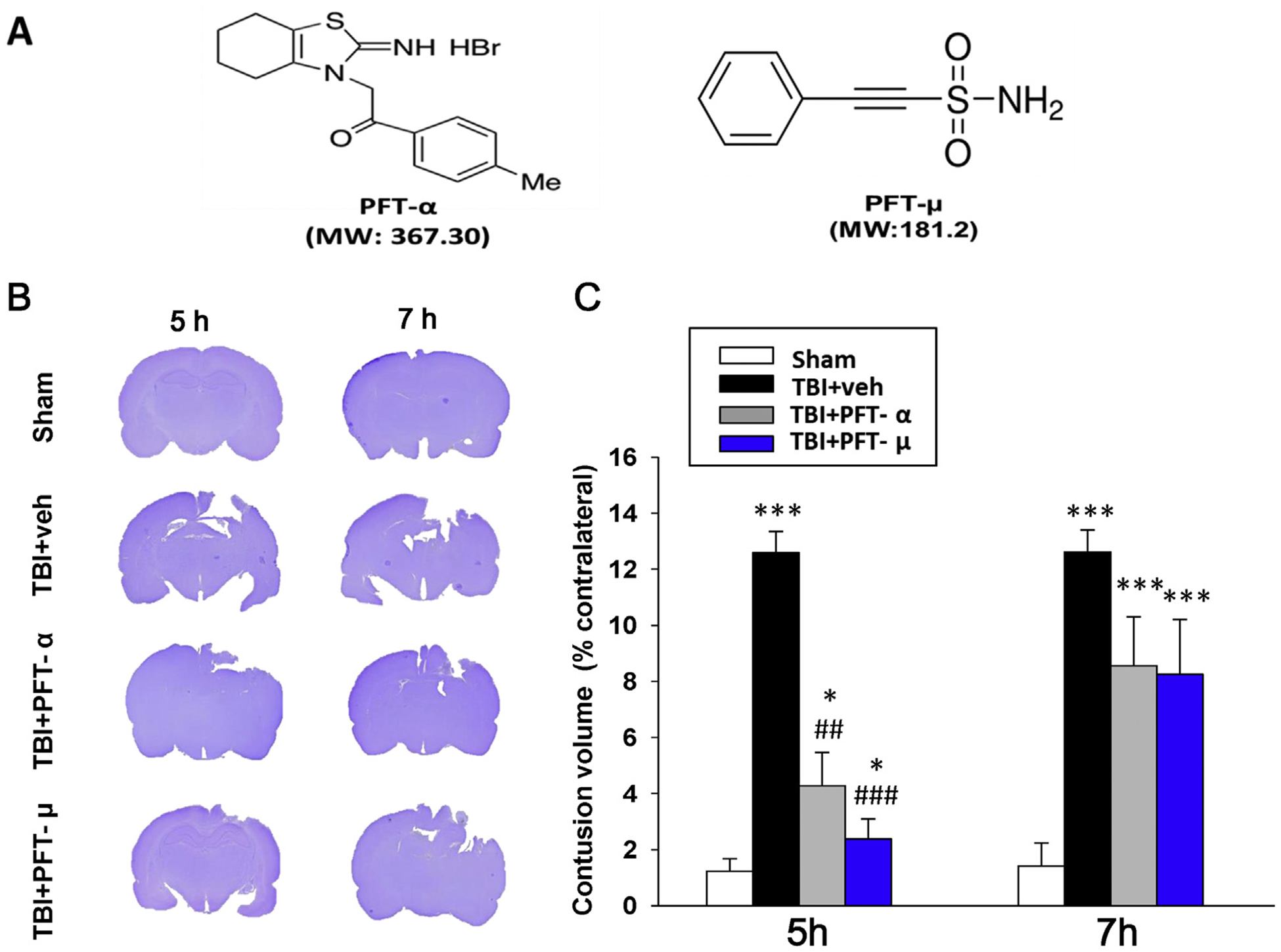
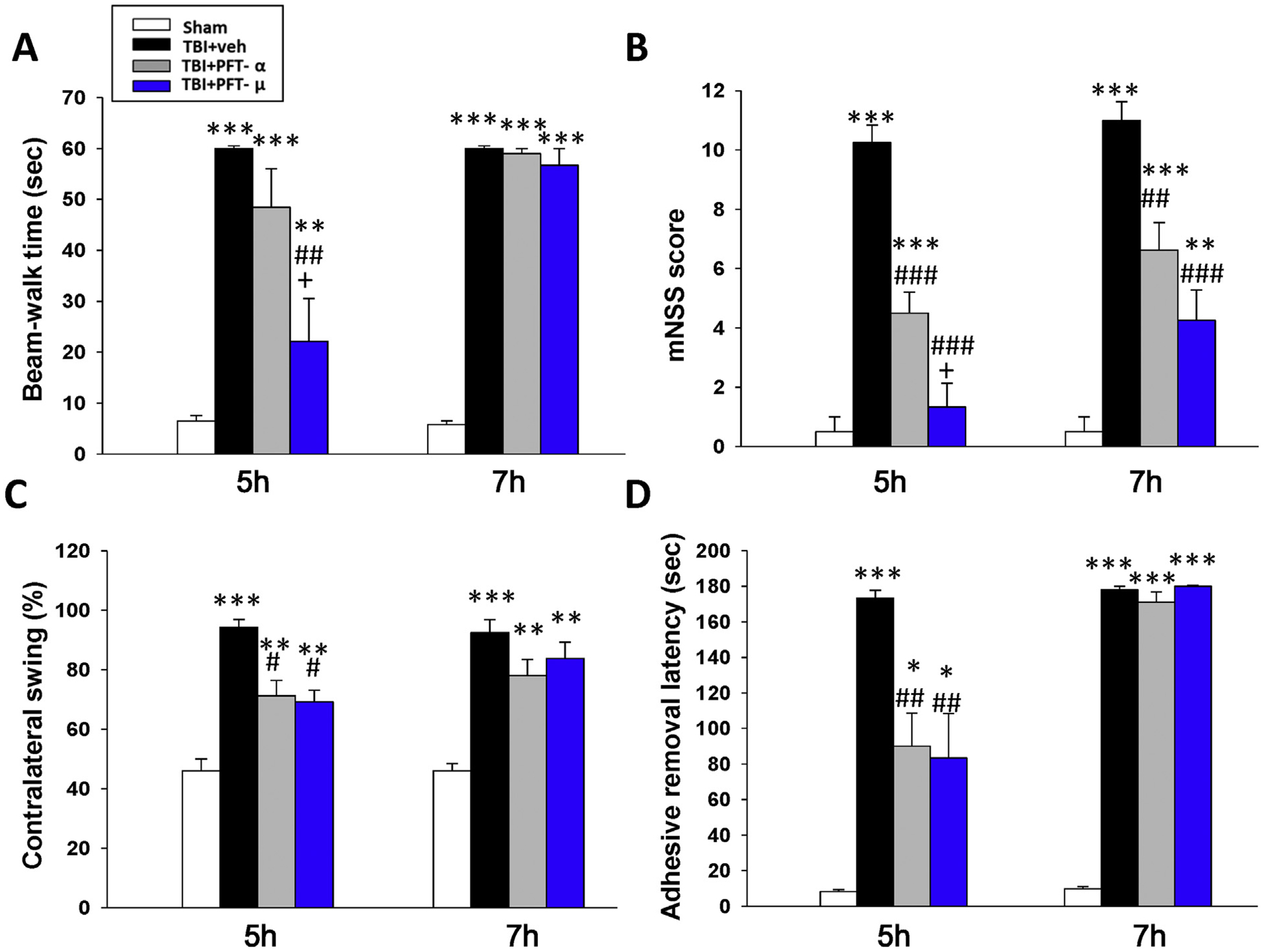
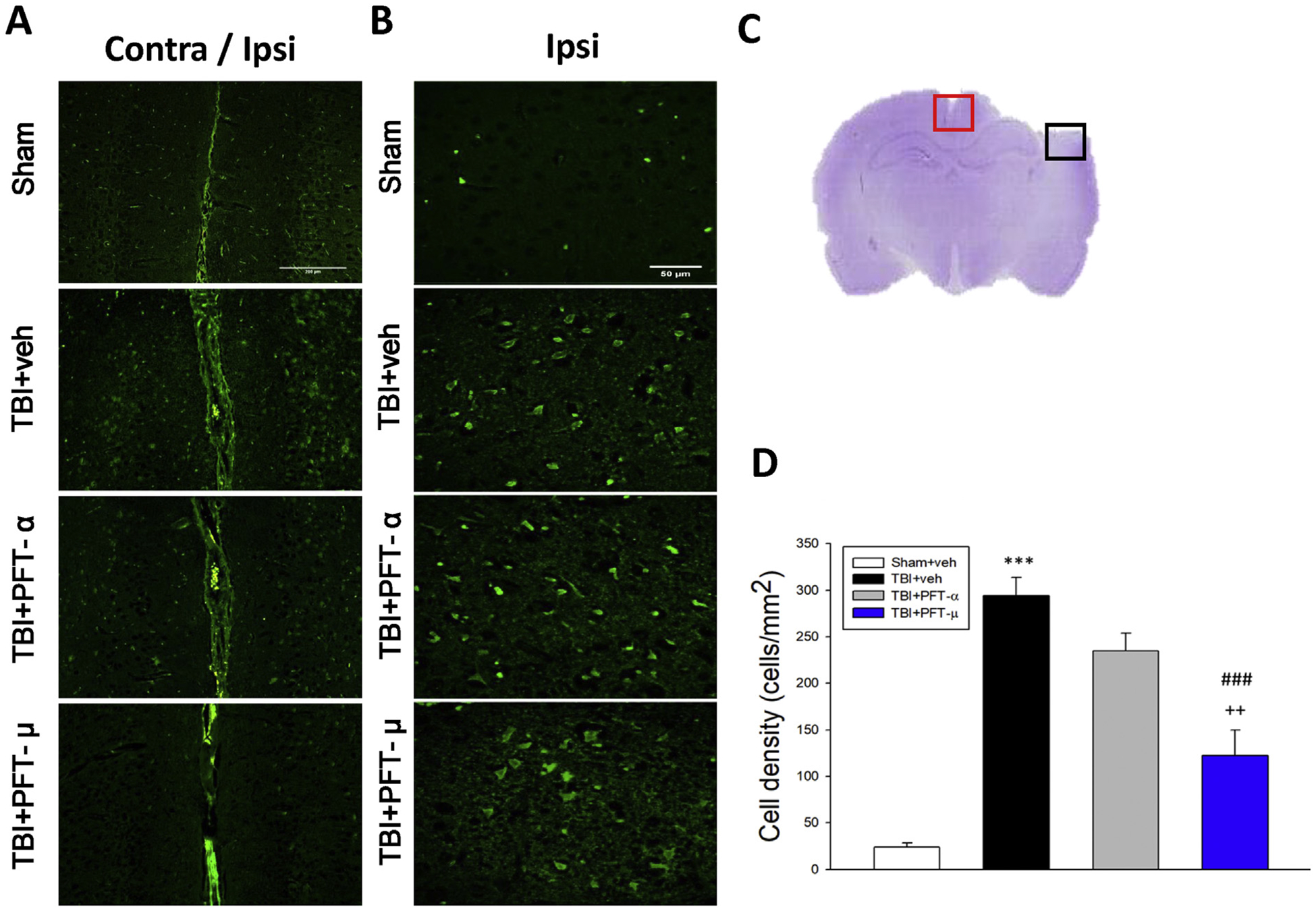
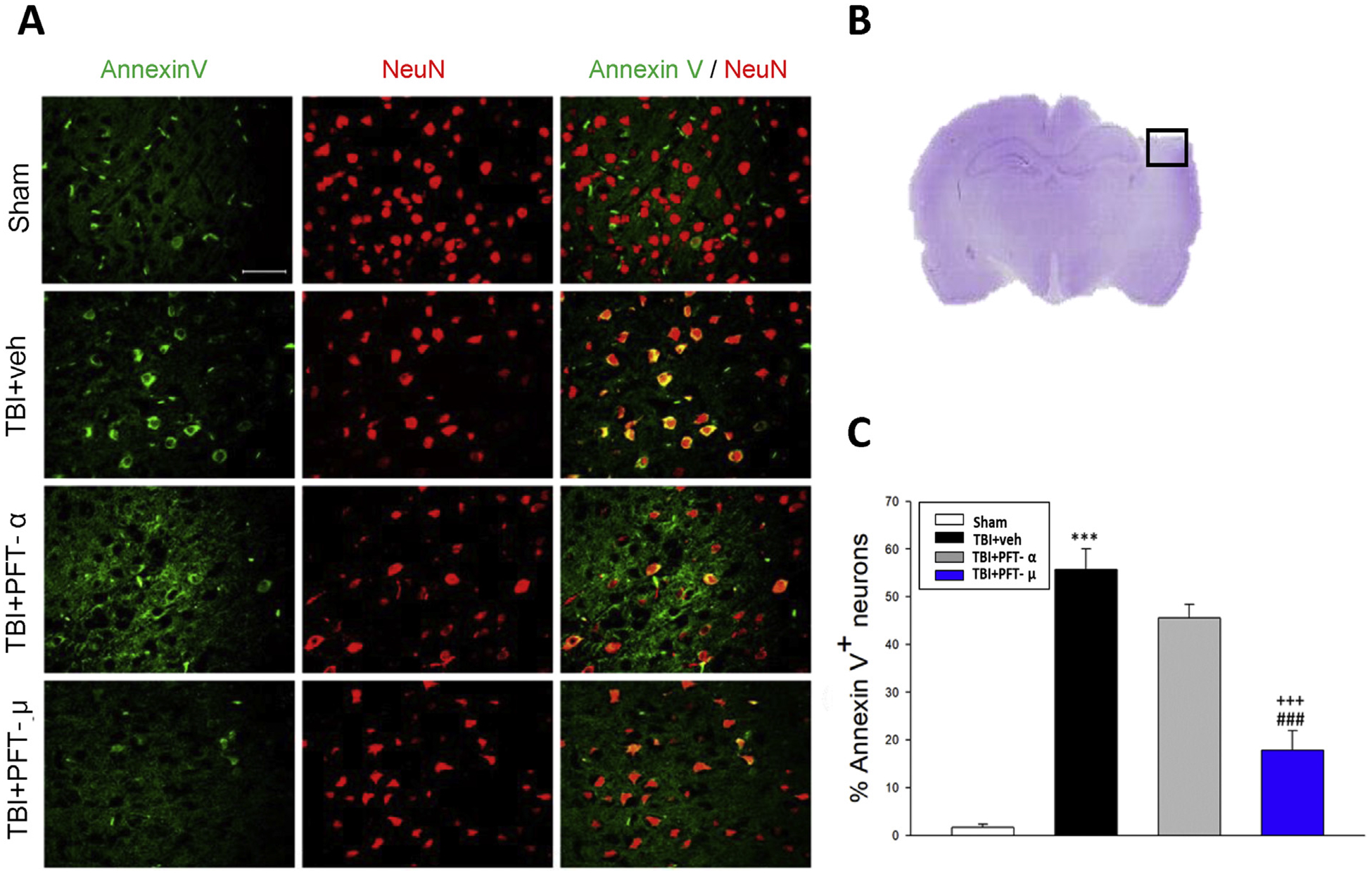
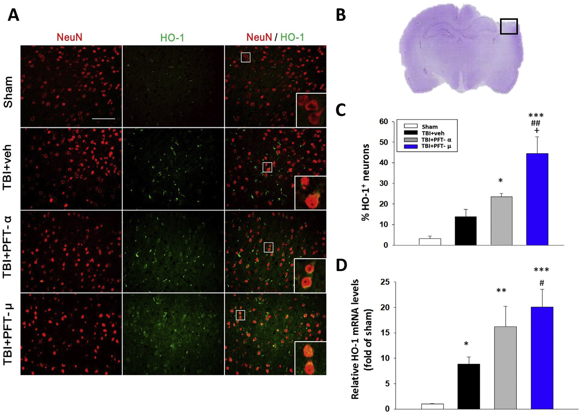
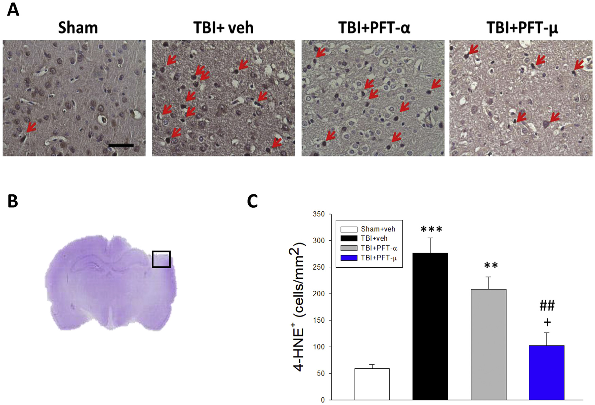
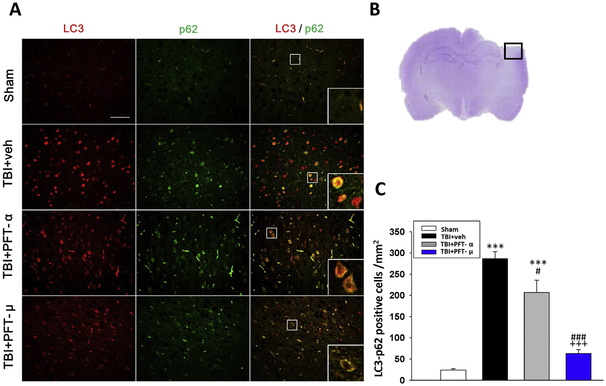
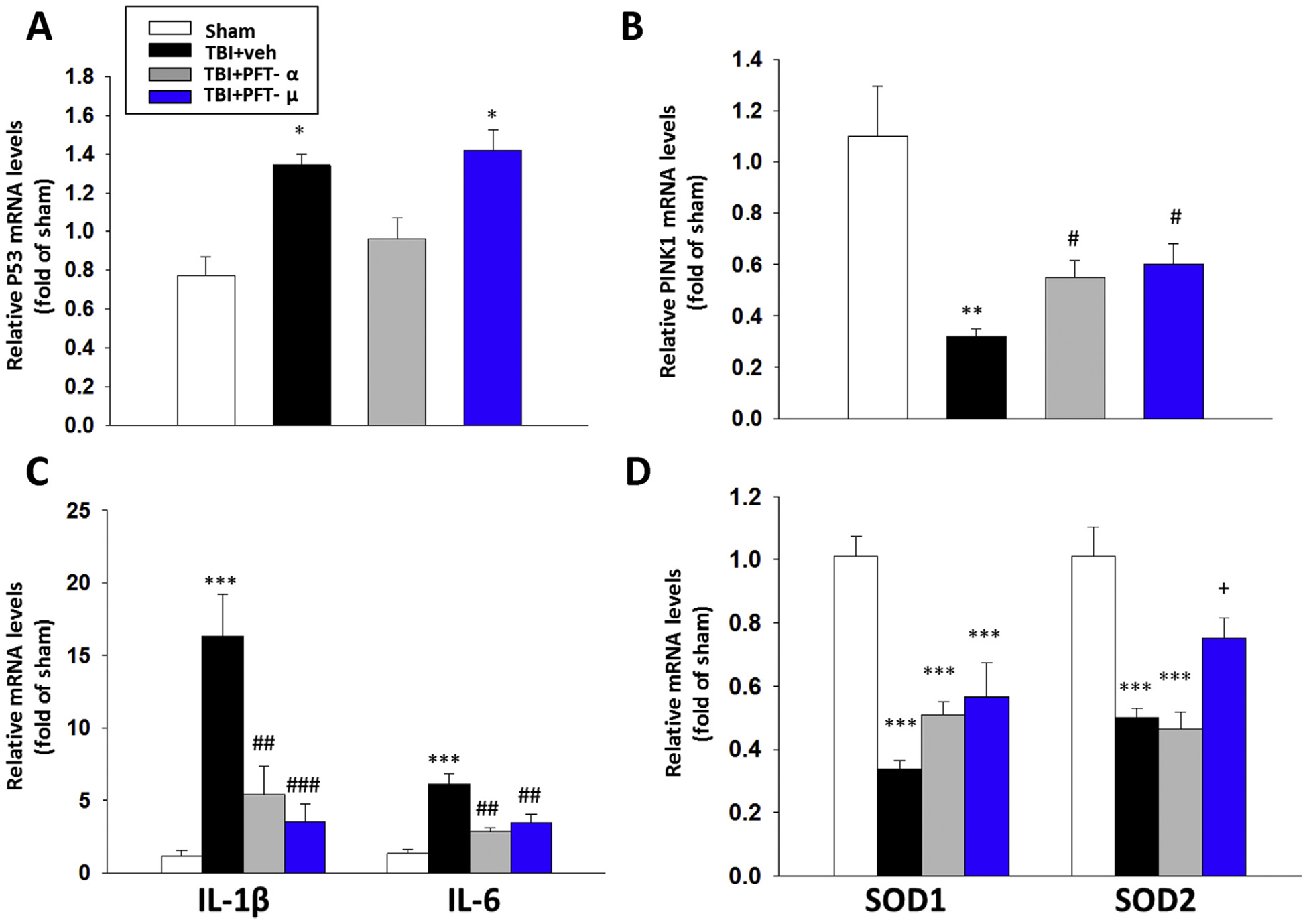
Traumatic brain injury (TBI) is one of the most common causes of death and disability worldwide. We investigated whether inhibition of p53 using pifithrin (PFT)-α or PFT-μ provides neuroprotective effects via p53 transcriptional dependent or -independent mechanisms, respectively. Sprague Dawley rats were subjected to controlled cortical impact TBI followed by the administration of PFTα or PFT-μ (2 mg/kg, i.v.) at 5 h after TBI. Brain contusion volume, as well as sensory and motor functions were evaluated at 24 h after TBI. TBI-induced impairments were mitigated by both PFT-α and PFT-μ. Fluoro-Jade C staining was used to label degenerating neurons within the TBI-induced cortical contusion region that, together with Annexin V positive neurons, were reduced by PFT-μ. Double immunofluorescence staining similarly demonstrated that PFT-μ significantly increased HO-1 positive neurons and mRNA expression in the cortical contusion region as well as decreased numbers of 4-hydroxynonenal (4HNE)-positive cells. Levels of mRNA encoding for p53, autophagy, mitophagy, anti-oxidant, anti-inflammatory related genes and proteins were measured by RT-qPCR and immunohistochemical staining, respectively. PFT-α, but not PFT-μ, significantly lowered p53 mRNA expression. Both PFT-α and PFT-μ lowered TBI-induced pro-inflammatory cytokines (IL-1β and IL-6) mRNA levels as well as TBI-induced autophagic marker localization (LC3 and p62). Finally, treatment with PFT-μ mitigated TBI-induced declines in mRNA levels of PINK-1 and SOD2. Our data suggest that both PFT-μ and PFT-α provide neuroprotective actions through regulation of oxidative stress, neuroinflammation, autophagy, and mitophagy mechanisms, and that PFT-μ, in particular, holds promise as a TBI treatment strategy.

Keywords: Autophagy; Mitophagy; Neuroinflammation; PFT-α; PFT-μ; Pifithrin analogs; Traumatic brain injury (TBI); p53.


Conflict of interest statement

Declaration of Competing Interest

The authors declare no conflicts of Interest.

Figures

Fig. 1.
Fig. 1.
Post-injury administration of PFT-α or PFT-μ at 5 h after TBI significantly reduced contusion volume at 24 h. (A) Structure and molecular weight (MW) of PFT-α and PFT-μ. (B) Coronal sections of sham, vehicle, and PFT-α/ PFT-μ 5 h and 7 h treated TBI rats were obtained and stained with cresyl violet at 24 h post injury. (C) The contusion volumes of TBI groups were not different from one another at 7 h, but at 5 h were significantly reduced by PFT-α or PFT-μ treatment compared to the TBI vehicle (veh) group. Data are expressed as means ± SEM. * P < .05, *** P < .001 versus sham group; ## P < .01, ### P < .001, versus TBI + veh group (n = 8 in TBI + veh and TBI + PFT-α group, n = 5 in sham and TBI + PFT-μ group).
Fig. 2.
Fig. 2.
Post-injury administration of PFT-α or PFT-μ given at 5 h improved functional outcomes, as revealed by multiple behavioral evaluations undertaken at 24 h post CCI or sham challenge. (A) Motor coordination measured by beam walking test. (B) Neurological function measured by mNSS. (C) Motor asymmetry measured by elevated body swing test (EBST). (D) Sensory-motor function measured by tactile adhesive removal test. Data represent the means ± SEM. *P < .05, **P < .01, ***P < .001 versus sham group; #P < .05, ##P < .01, ###P < .001 versus TBI + vehicle (veh) group; +P < .05 versus TBI + PFT-α group. (n = 8 in TBI + veh and TBI + PFT-α group, n = 5 in sham and TBI + PFT-μ group). Note that when PFT treatment was administered at 7 h post injury, it proved much less effective.
Fig. 3.
Fig. 3.
Post-injury administration of PFT-μ at 5 h after TBI significantly decreased FJC-positive cells at 24 h. FJC-stained (A) contralateral and ipsilateral (Bar = 200 μm) (B) ipsilateral cortex (Bar = 50 μm) regions of interest in sham, TBI + veh, TBI + PFT-α or TBI + PFT-μ group. (C) A representative HE-stained coronal section showing the area as indicated by the red (for both contralateral and ipsilateral cortex) and black (only ipsilateral cortex) square box to compare the fluorescent signals across the 4 groups of rats. (D) There was a significant decrease in the number of FJC-positive cells in the TBI + PFT-μ group. Data are expressed as means ± SEM. The total number of FJC-positive cells was expressed as the mean number per field of view (n = 5 per group). ***P < .001 versus sham group; ###P < .001 versus TBI + vehicle (veh) group; ++P < .01 versus TBI + PFT- α group.
Fig. 4.
Fig. 4.
Post-injury administration of PFT-μ at 5 h after TBI decreased annexin V positive neurons in the cortical contusion region at 24 h. (A) Co-immunohistochemistry of annexin V and NeuN in the cortical region. Annexin V (a marker of apoptosis) immunoreactivity is shown in green, and NeuN (a cell marker for neurons) is shown in red. Yellow labelling indicates colocalization. (B) A representative HE-stained coronal section showing the area as indicated by the black (ipsilateral cortex) square box to compare the fluorescent signals across the 4 groups of rats. (C) There was a significant decrease in the number of Annexin V positive neurons in the TBI + PFT-μ group. Data are expressed as means ± SEM (n = 5 per group). ***P < .001, compared with the sham group; ###P < .001, versus the TBI + veh group; +++P < .001 versus the TBI + PFT-α group. Scale bar = 50 um.
Fig. 5.
Fig. 5.
Post-injury administration of PFT-α and, in particular, PFT-μ at 5 h after TBI elevated HO-1 positive neurons and mRNA expression in the cortical contusion region at 24 h. (A) Co-immunohistochemistry of HO-1 and NeuN in cortical brain tissue. (B) A representative HE-stained coronal section showing the area as indicated by the black (ipsilateral cortex) square box to compare the fluorescent signals across the 4 groups of rats. (C) There was a significant increase of HO-1 positive neurons in the TBI + PFT-μ group beyond that induced in the TBI + vehicle group. (D) The mRNA levels of HO-1 were analyzed by RT-PCR. Data are expressed as means ± SEM (n = 5 per group). *P < .05, **P < .01, ***P < .001, compared with the sham group; #P < .05, ##P < .01, compared with the TBI + veh group; +P < .05, compared with the TBI + PFT-α group. Scale bar = 100 μm.
Fig. 6.
Fig. 6.
Post-injury administration of PFT-μ, but not PFT-α, at 5 h after TBI decreased 4-HNE positive cells in the cortical contusion region at 24 h. (A) Representative photomicrographs showing the results of immunohistochemical staining with anti-4HNE in 4 groups. Red arrows indicate 4-HNE-positive cells (brown colour). (B) A representative HE-stained coronal section showing the area as indicated by the black (ipsilateral cortex) square box to compare the fluorescent signals across the 4 groups of rats. (C) There was a significant decrease in the number of 4-HNE-positive cells in the TBI + PFT-μ group. The total number of 4-HNE-positive cells was expressed as the mean number of cells/mm2. Mean ± SEM. **P < .01, ***P < .001, compared with the sham group. ##P < .01, compared with the TBI + veh group. +P < .05, compared with the TBI + PFT-α group. (Scale bar = 50 μm; n = 5 per group).
Fig. 7.
Fig. 7.
Post-injury administration of PFT-μ at 5 h after TBI decreases LC3/p62 positive cells in the cortical contusion region at 24 h. (A) Co-immunohistochemistry of LC3 and p62 in cortical brain tissue. LC3: green, p62: red, and yellow labelling indicating colocalization (Bar = 100 μm, 0.259 mm2). Inserts: Magnified colocalization of LC3 (red) and p62 (green) cells at a greater magnification. (B) A representative HE-stained coronal section showing the area as indicated by the black (ipsilateral cortex) square box to compare the fluorescent signals across the 4 groups of rats. (C) A significant decrease in LC3/p62 positive cells was evident in the TBI + PFT-μ group. The total number of LC3/p62-positive cells was expressed as the mean number per field of view in cortex. Data represent mean ± S.E.M. (n = 5 per group). **P < .01, ***P < .001 compared to the sham group; #P < .05, ### P < .001 compared to the TBI + veh group; +++ P < .001 compared to the TBI + PFT-α group.
Fig. 8.
Fig. 8.
Treatment with PFT-μ at 5 h after TBI significantly mitigated TBI-induced reductions in PINK-1, and SOD2, and inhibited TBI-mediated rises in IL-1β, IL-6 mRNA expression in the cortical contusion region at 24 h. The mRNA levels of (A) p53, (B) PINK1, (C) IL-1β and IL-6, and (D) SOD1 and SOD2 across the four animal groups were evaluated by RT-PCR. PFT-μ significantly increased PINK1 and SOD2 mRNA expression, and decreased IL1β and IL-6 mRNA compared with TBI + vehicle-treated rats. Notably, PFT-μ did not change the level of p53 gene expression in accord with its transcriptional independent mechanism of action. Data are expressed as means ± SEM (n = 5 per group). * P < .05, **P < .01, ***P < .001 vs. sham; #P < .05, ##P < .01, ###P < .001 vs. TBI + veh group. +P < .05 vs. TBI + PFT-α group.

References

    1. Alder J, Fujioka W, Lifshitz J, Crockett DP, Thakker-Varia S, 2011. Lateral fluid percussion: model of traumatic brain injury in mice. J. Vis. Exp 3063. - PMC - PubMed
    1. Alluri H, Wiggins-Dohlvik K, Davis ML, Huang JH, Tharakan B, 2015. October Blood-brain barrier dysfunction following traumatic brain injury. Metab. Brain Dis 30 (5), 1093–1104. 10.1007/s11011-015-9651-7. - DOI - PubMed
    1. Arango D, Corner GA, Wadler S, Catalano PJ, 2001. Augenlicht, L.H. C-myc/p53 interaction determines sensitivity of human colon carcinoma cells to 5-fluorouracil in vitro and in vivo. Cancer Res. 61, 4910–4915. - PubMed
    1. Baratz R, Tweedie D, Wang JY, Rubovitch V, Luo W, Hoffer BJ, Greig NH, Pick CG, 2015. Transiently lowering tumor necrosis factor-α synthesis ameliorates neuronal cell loss and cognitive impairments induced by minimal traumatic brain injury in mice. J. Neuroinflammation 12. - PMC - PubMed
    1. Barkhoudarian G, Hovda DA, Giza CC, 2011. The molecular pathophysiology of concussive brain injury. Clin. Sports Med 30, 33–48. - PubMed

Publication types

MeSH terms