Review
. 2019 Nov 26;8(12):1515.
doi: 10.3390/cells8121515.

Histamine-Releasing Factor, a New Therapeutic Target in Allergic Diseases

Affiliations
Review

Histamine-Releasing Factor, a New Therapeutic Target in Allergic Diseases

Yu Kawakami et al. Cells. .

Abstract

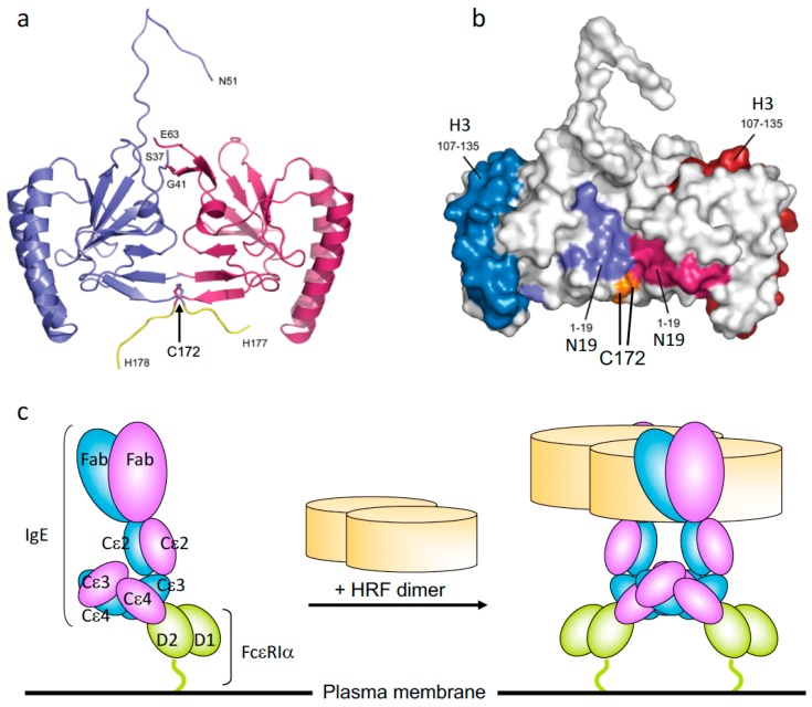
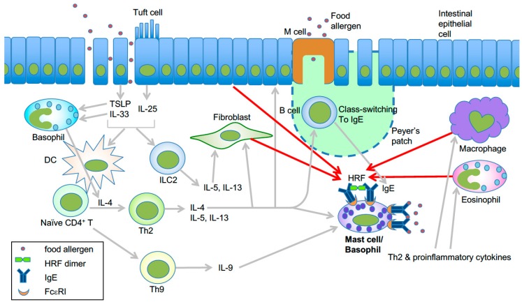
Histamine-releasing activities on human basophils have been studied as potential allergy-causing agents for four decades. An IgE-dependent histamine-releasing factor (HRF) was recently shown to interact with a subset of immunoglobulins. Peptides or recombinant proteins that block the interactions between HRF and IgE have emerged as promising anti-allergic therapeutics, as administration of them prevented or ameliorated type 2 inflammation in animal models of allergic diseases such as asthma and food allergy. Basic and clinical studies support the notion that HRF amplifies IgE-mediated activation of mast cells and basophils. We discuss how secreted HRF promotes allergic inflammation in vitro and in vivo complex disease settings.

Keywords: FcεRI; HRF; IgE; allergy; basophils; mast cells; translationally controlled tumor protein (TCTP).


Conflict of interest statement

The authors declare no conflict of interest.

Figures

Figure 1
Figure 1
The crystal structure of histamine-releasing factor (HRF) dimer and HRF dimer/IgE-mediated FcεRI crosslinking. (a) Overall structure of a human HRF dimer. The two molecules of the asymmetric unit are colored blue and pink. The C-terminal tag is colored yellow, and the positions of C-terminal residues and residues adjacent to the disordered loop are indicated. (b) The two monomers of the HRF dimer are colored white and Cys172 is colored orange. For the first monomer, the two IgE binding sites, mapped to residues Met1–Lys19 (N19), and Arg107–Ile135 (H3), are colored light blue and dark blue, respectively. For the second monomer, residues 1-19 (N19) and 107-135 (H3) are colored light and dark pink, respectively. (c) Model for HRF dimer/IgE-mediated FcεRI crosslinking. IgE binds FcεRI α chain via the interaction between IgE–Cε3 and FcεRIα–D2 domains. One HRF molecule can bind one (this version depicted) or two molecules of IgE via interactions with the N19 and H3 regions of HRF. After binding of an HRF dimer, two (this version depicted) or four FcεRI α chain-nucleated complexes will be formed (Right). The cytoplasmic portion of FcεRI α as well as β and γ chains of FcεRI are omitted for clarity.
Figure 2
Figure 2
Model of HRF-mediated amplification of type 2 inflammation in food allergy. Epithelial damage or inflammation in the gut promotes increased entry of food allergens and secretion of the epithelial cytokines TSLP, IL-25, and IL-33 [44]. These cytokines initiate a Th2-skewed immune response. TSLP can enhance OX40L expression in dendritic cells, which induce Th2 cell differentiation of naïve CD4+ T cells [45]. IL-25 secreted by tuft cells may help the expansion of type 2 innate lymphoid cells (ILC2) [46]. Th2 cells along with ILC2 cells promote the Th2 cell-mediated immune response, which includes IgE class switch recombination in B cells, eosinophil accumulation, and mastocytosis. IL-9 promotes the expansion of IL-9-producing mucosal mast cells [47]. Basophils are also required for production of antigen-specific IgE as well as oral allergen-induced food allergy during sensitization [48,49] and allergen challenge phases [50]. IL-4 derived from basophils stimulated by cytokines such as IL-33 seems to be required for Th2 cell differentiation [51], and IL-4 promotes intestinal mast cell accumulation and activation [52]. HRF dimer/oligomers secreted from several types of cells amplify intestinal inflammation by enhancing antigen/IgE-mediated activation of mast cells and basophils [30]. This is likely due to increased HRF secretion by several types of cells in response to Th2, proinflammatory and epithelial cytokines. Modified from ref. 66 with permission from the journal Allergy.

References

    1. Metzger H. Transmembrane signaling: The joy of aggregation. J. Immunol. 1992;149:1477–1487. - PubMed
    1. Mukai K., Tsai M., Saito H., Galli S.J. Mast cells as sources of cytokines, chemokines, and growth factors. Immunol. Rev. 2018;282:121–150. doi: 10.1111/imr.12634. - DOI - PMC - PubMed
    1. Galli S.J., Tsai M. IgE and mast cells in allergic disease. Nat. Med. 2012;18:693–704. doi: 10.1038/nm.2755. - DOI - PMC - PubMed
    1. MacDonald S.M., Lichtenstein L.M. Histamine-releasing factors and heterogeneity of IgE. Springer Semin. Immunopathol. 1990;12:415–428. doi: 10.1007/BF00225327. - DOI - PubMed
    1. Kuna P., Reddigari S.R., Rucinski D., Oppenheim J.J., Kaplan A.P. Monocyte chemotactic and activating factor is a potent histamine-releasing factor for human basophils. J. Exp. Med. 1992;175:489–493. doi: 10.1084/jem.175.2.489. - DOI - PMC - PubMed

Publication types

MeSH terms