. 2019 Nov 26;11(12):1095.
doi: 10.3390/v11121095.

Insights into Innate Sensing of Prototype Foamy Viruses in Myeloid Cells

Affiliations

Insights into Innate Sensing of Prototype Foamy Viruses in Myeloid Cells

Maïwenn Bergez et al. Viruses. .

Abstract

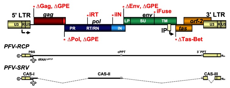
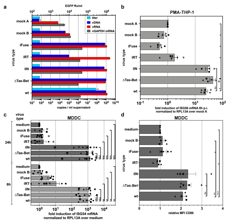
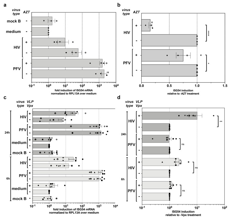
Foamy viruses (FVs) belong to the Spumaretrovirinae subfamily of retroviruses and are characterized by unique features in their replication strategy. This includes a reverse transcription (RTr) step of the packaged RNA genome late in replication, resulting in the release of particles with a fraction of them already containing an infectious viral DNA (vDNA) genome. Little is known about the immune responses against FVs in their hosts, which control infection and may be responsible for their apparent apathogenic nature. We studied the interaction of FVs with the innate immune system in myeloid cells, and characterized the viral pathogen-associated molecular patterns (PAMPs) and the cellular pattern recognition receptors and sensing pathways involved. Upon cytoplasmic access, full-length but not minimal vector genome containing FVs with active reverse transcriptase, induced an efficient innate immune response in various myeloid cells. It was dependent on cellular cGAS and STING and largely unaffected by RTr inhibition during viral entry. This suggests that RTr products, which are generated during FV morphogenesis in infected cells, and are therefore already present in FV particles taken up by immune cells, are the main PAMPs of FVs with full-length genomes sensed in a cGAS and STING-dependent manner by the innate immune system in host cells of the myeloid lineage.

Keywords: STING; cGAS; foamy virus; innate sensing; retrovirus; spumavirus.


Conflict of interest statement

The authors declare no conflict of interest. The funders had no role in the design of the study; in the collection, analyses, or interpretation of data; in the writing of the manuscript, or in the decision to publish the results.

Figures

Figure A1
Figure A1
PFV genome organization and viral expression systems used. Schematic outline of the wild type (wt) PFV proviral genome structure with gag, pol, and env ORFs. The locations of the point mutations for the individual proviral mutants resulting in variants with enzymatically inactive integrase (iIN) or reverse transcriptase (iRT); fusion-deficient Env (iFuse); or deficiencies in the translation of, Tas (∆Tas-Bet); Gag (∆Gag); Pol (∆Pol); Env (∆Env); or Gag, Pol, and Env (∆GPE), due to inactivation of the respective translation initiation sites. PFV-RCP supernatants contain full-length viral genomic RNA (vgRNA) whereas PFV-SRV supernatants harbor vgRNA comprising the minimal essential cis-acting viral sequences (CAS). LTR: long terminal repeat; U3: unique 3′ LTR region; U5: unique 5′ LTR region; R: repeat LTR region; PR: protease; RT/RN: reverse transcriptase—RNase H; IN: integrase; LP leader peptide; SU: surface subunit; TM: transmembrane subunit; IP: internal promoter; ©: cap; An: poly-A tail; PBS: primer binding site; 3′ PPT: 3′ poly purine tract; cPPT: central PPT.
Figure 1
Figure 1
PFV-mediated ISG induction in myeloid cells. (a,b) Kinetics of ISG56/ISG54 induction in PMA-differentiated THP-1 wild type cells incubated with different amounts of wild type PFV-RCP (a, MOI 0.2) as well as pUC19 (mock A) mock supernatants. ISG mRNA levels normalized for RPL13A mRNA levels were determined by qPCR at the indicated time points post exposure. Means ± SDs of ISG56 (n = 4; a) or ISG54 (n = 4; b) induction relative to mock A treatment are shown. (c,d) Primary human MDDC (c) or MDM (d) were incubated with wild type PFV-RCP (PFV; MOI 0.25), ∆GPE (mock B) mock supernatant, or medium (medium) for 6, 12, or 24 h, as indicated. Means ± SEMs, plus individual data points, of ISG54 (n = 5–8) induction normalized to RPL13A relative to medium treatment are shown. Mixed-effects analysis with Holm–Sidak’s multiple-comparisons test was used to assess significance. * p < 0.05; ** p < 0.01; *** p < 0.001; **** p < 0.0001; ns: not significant (p ≥ 0.05).
Figure 2
Figure 2
cGAS and STING-mediated sensing of PFV-associated reverse transcription products. PMA-differentiated THP-1 wild type cells and KO variants with deficiencies in components of various innate sensing pathways, as indicated, were incubated with wild type PFV-RCP (MOI 0.2) or pUC19 (mock A) mock supernatants. ISG mRNA levels normalized for RPL13A mRNA levels were determined by qPCR at the indicated time points post exposure. Means ± SEM, plus individual data points, of ISG56 (n = 3) induction normalized to RPL13A relative to mock A treatment are shown. Two-way ANOVA with Tukey’s multiple-comparisons test was used to assess significance. * p < 0.05; ** p < 0.01; *** p < 0.001; **** p < 0.0001; ns: not significant (p ≥ 0.05).
Figure 3
Figure 3
Differential ISG induction profiles of PFV mutants (described in detail in Figure A1 and Material and Methods) varying in their protein and nucleic acid composition. (a) Particle-associated nucleic acids extracted from viral particles pelleted by ultracentrifugation of virus supernatants used in (bd) were analyzed by qPCR to quantify the particle-associated viral (vDNA: viral DNA; vRNA: viral RNA) and cellular (vGAPDH mRNA) nucleic acid composition. Mean copy numbers ± SDs per mL supernatant determined from duplicates are shown. (b,c) ISG induction profile of PMA-differentiated THP-1 wild type cells (b) or MDDCs (c) incubated with identical amounts of wild type PFV-RCP (MOI 0.1 THP-1; MOI 0.25 MDDCs) supernatants, variants thereof, and pUC19 (mock A) and ∆GPE (mock B) mock supernatants, or medium, as indicated, for 8 h and 24 h. Means ± SEMs, plus individual data points, of ISG56 (n = 6) or ISG54 (n = 5–7) mRNA induction normalized to RPL13A mRNA relative to mock A or medium treatment are shown. One-way ANOVA with Tukey’s multiple-comparisons test (b) or mixed-effects analysis with Tukey’s multiple-comparisons test (c) was used to assess significance. (d) CD86 cell surface expression profile of MDDCs 24 h post exposure to wild type PFV-RCP (MOI 0.25) supernatants and variants thereof as indicated. Means ± SEMs (n = 5), plus individual data points, relative to medium treatment are shown. Mixed-effects analysis with Tukey’s multiple-comparisons test was used to assess significance. * p < 0.05; ** p < 0.01; *** p < 0.001; **** p < 0.0001; ns: not significant (p ≥ 0.05).
Figure 4
Figure 4
Influence of RTr inhibition and VLP-Vpx treatment on PFV-mediated ISG induction during target-cell entry. (a,b) MDDCs were incubated with wild type PFV-RCP (MOI 0.25), HIV-1 GFP (MOI 2), and VLP-Vpx or ∆GPE (mock B) mock supernatants in the absence or presence of AZT (100 µM) as indicated. ISG54 mRNA levels normalized for RPL13A mRNA levels were determined by qPCR at 24 h post exposure. (a) Mean values ± SEMs, plus individual data points of ISG54 (n = 3–4) induction relative to medium incubated samples are shown. (b) Mean values ± SEMs, plus individual data points of ISG54 (n = 4) induction relative to the respective sample incubated with the same virus type without AZT addition are shown. One-way ANOVA with Sidak’s multiple-comparisons test was used to assess significance. (c,d) MDDCs were incubated with wild type PFV-RCP (MOI 0.25), HIV-1 GFP (MOI 2) supernatants, or ∆GPE (mock B) mock supernatants in the absence or presence of VLP-Vpx as indicated. ISG54 mRNA levels normalized for RPL13A mRNA levels were determined by qPCR at 6 and 24 h post exposure. (c) Mean values ± SEMs, plus individual data points, of ISG54 (n = 7) induction relative to medium incubated samples are shown. (d) Mean values ± SEMs, plus individual data points, of ISG54 (n = 7) induction relative to the respective sample incubated with the same virus type without VLP-Vpx addition are shown. One-way ANOVA with Sidak’s multiple-comparisons test was used to assess significance. * p < 0.05; ** p < 0.01; *** p < 0.001; **** p < 0.0001; ns: not significant (p ≥ 0.05).
Figure 5
Figure 5
Differential ISG induction profile of single round PFV vector particles harboring full-length or minimal viral genomes. (a) PMA-differentiated THP-1 wild type cells were incubated with the indicated relative amounts of wild type PFV-RCP (MOI 0.3) supernatants and variants thereof, or the different PFV-SRV supernatants with variable Pol content (µg Pol packaging plasmid used for supernatant production is indicated; MOI 3 at 2.5 µg Pol) or supernatant from 293T cells transfected with pUC19 (mock A). ISG56 mRNA levels normalized for RPL13A mRNA levels were determined by qPCR at 8 h post exposure. Mean values ± SEMs, plus individual data points of ISG56 (n = 1–3) induction relative to mock A treatment are shown. (b,c) PFV supernatants characteristics. (b) Particle protein composition. Western blot analysis of protein composition of viral particles pelleted by ultracentrifugation of virus supernatants used in panel A employing PFV Gag (α-PFV Gag) and PFV Env LP (α-PFV Env LP) specific polyclonal antisera. The identity of individual protein bands is indicated to the left, the molecular weight to the right. (c) Particle nucleic acid composition. Particle-associated nucleic acids extracted from viral particles pelleted by ultracentrifugation of virus supernatants used in panel A were analyzed by qPCR to quantify the particle-associated viral (vDNA: viral DNA; vRNA: viral RNA) and cellular (vGAPDH mRNA) nucleic acid composition. Mean copy numbers ± SDs per mL supernatant determined from duplicates are shown. Viral titers were determined for PFV-SRV supernatants by EGFP reporter gene transfer assay on HT1080 cells and for PFV-RCP supernatants by Tas-dependent EGFP reporter gene induction assays on HT1080 PLNE cells. Mean titers ± SDs per mL supernatant determined from technical duplicates are shown.

References

    1. Khan A.S., Bodem J., Buseyne F., Gessain A., Johnson W., Kuhn J.H., Kuzmak J., Lindemann D., Linial M.L., Lochelt M., et al. Spumaretroviruses: Updated taxonomy and nomenclature. Virology. 2018;516:158–164. doi: 10.1016/j.virol.2017.12.035. - DOI - PMC - PubMed
    1. Rethwilm A., Lindemann D. Foamy viruses. In: Knipe D.M., Howley P.M., editors. Fields Virology. 6th ed. Volume 2. Lippincott Williams & Wilkins, a Wolters Kluwer business; Philadelphia, PA, USA: 2013. pp. 1613–1632.
    1. Lindemann D., Rethwilm A. Foamy virus biology and its application for vector development. Viruses. 2011;3:561–585. doi: 10.3390/v3050561. - DOI - PMC - PubMed
    1. Delelis O., Saib A., Sonigo P. Biphasic DNA synthesis in spumaviruses. J. Virol. 2003;77:8141–8146. doi: 10.1128/JVI.77.14.8141-8146.2003. - DOI - PMC - PubMed
    1. Moebes A., Enssle J., Bieniasz P.D., Heinkelein M., Lindemann D., Bock M., McClure M.O., Rethwilm A. Human foamy virus reverse transcription that occurs late in the viral replication cycle. J. Virol. 1997;71:7305–7311. - PMC - PubMed

Publication types

Substances