

MDM2 inhibitor APG-115 synergizes with PD-1 blockade through enhancing antitumor immunity in the tumor microenvironment
- PMID: 31779710
- PMCID: PMC6883539
- DOI: 10.1186/s40425-019-0750-6
MDM2 inhibitor APG-115 synergizes with PD-1 blockade through enhancing antitumor immunity in the tumor microenvironment
Abstract
Background: Programmed death-1 (PD-1) immune checkpoint blockade has achieved clinical successes in cancer therapy. However, the response rate of anti-PD-1 agents remains low. Additionally, a subpopulation of patients developed hyperprogressive disease upon PD-1 blockade therapy. Combination therapy with targeted agents may improve immunotherapy. Recent studies show that p53 activation in the myeloid linage suppresses alternative (M2) macrophage polarization, and attenuates tumor development and invasion, leading to the hypothesis that p53 activation may augment antitumor immunity elicited by anti-PD-1 therapy.
Method: Using APG-115 that is a MDM2 antagonist in clinical development as a pharmacological p53 activator, we investigated the role of p53 in immune modulation and combination therapy with PD-1 blockade.
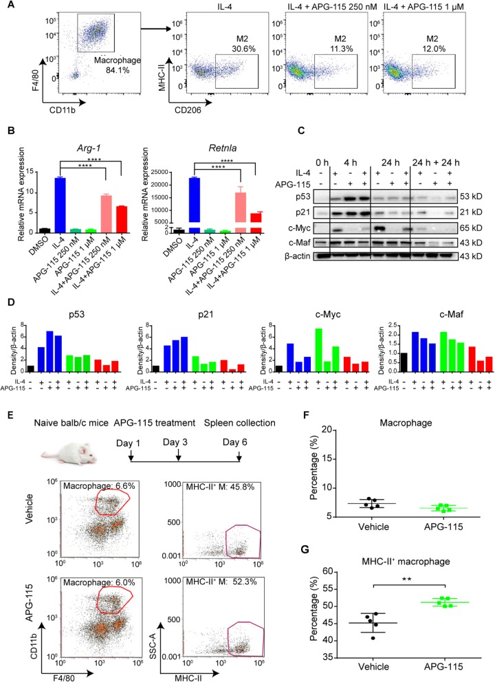
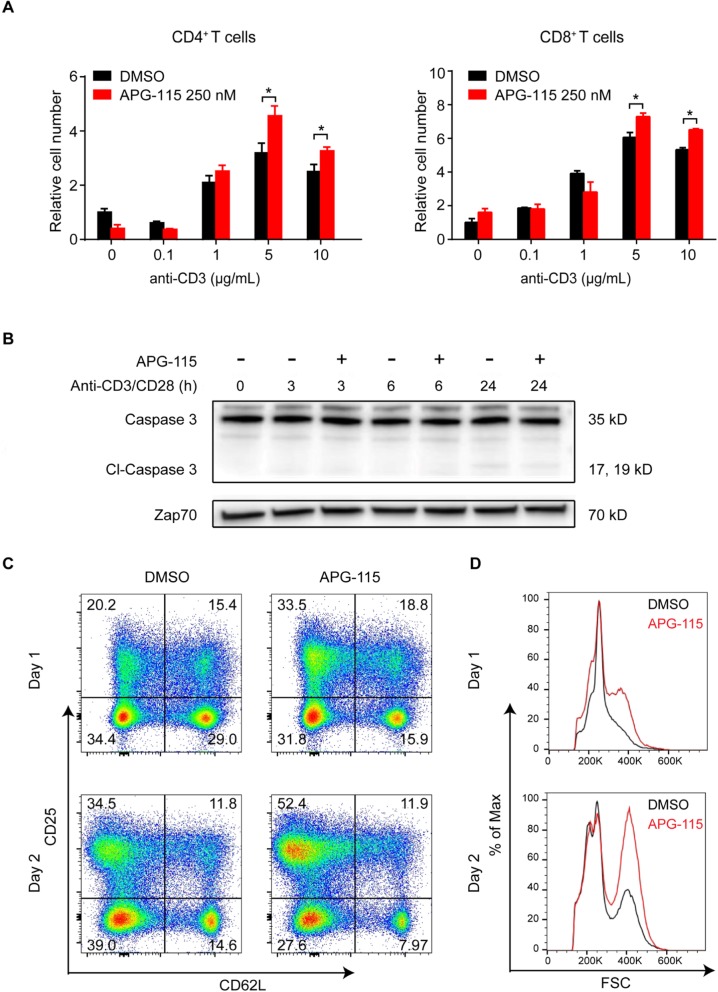
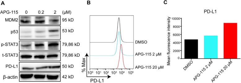
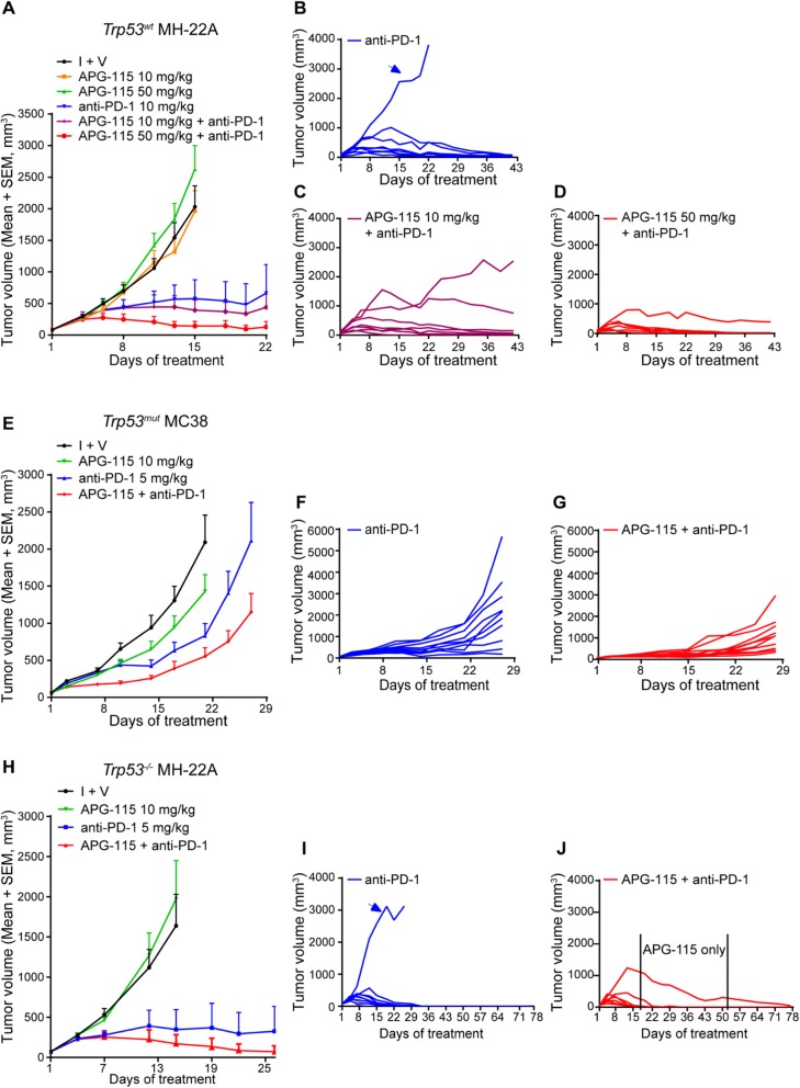
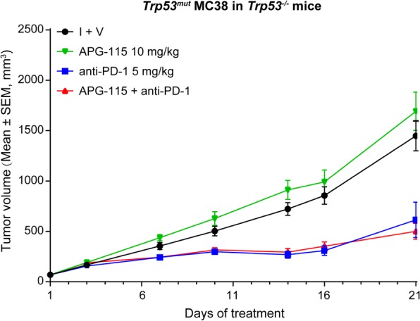
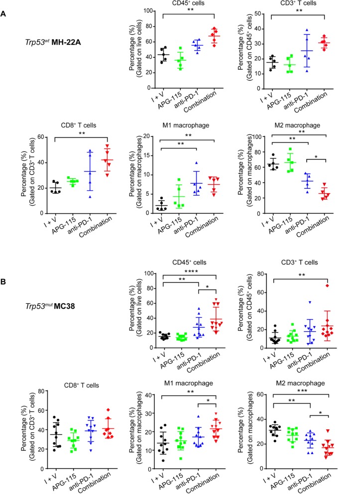
Results: In vitro treatment of bone marrow-derived macrophages with APG-115 resulted in activation of p53 and p21, and a decrease in immunosuppressive M2 macrophage population through downregulation of c-Myc and c-Maf. Increased proinflammatory M1 macrophage polarization was observed in the spleen from mice treated with APG-115. Additionally, APG-115 has co-stimulatory activity in T cells and increases PD-L1 expression in tumor cells. In vivo, APG-115 plus anti-PD-1 combination therapy resulted in enhanced antitumor activity in Trp53wt, Trp53mut, and Trp53-deficient (Trp53-/-) syngeneic tumor models. Importantly, such enhanced activity was abolished in a syngeneic tumor model established in Trp53 knockout mice. Despite differential changes in tumor-infiltrating leukocytes (TILs), including the increases in infiltrated cytotoxic CD8+ T cells in Trp53wt tumors and M1 macrophages in Trp53mut tumors, a decrease in the proportion of M2 macrophages consistently occurred in both Trp53wt and Trp53mut tumors upon combination treatment.
Conclusion: Our results demonstrate that p53 activation mediated by APG-115 promotes antitumor immunity in the tumor microenvironment (TME) regardless of the Trp53 status of tumors per se. Instead, such an effect depends on p53 activation in Trp53 wild-type immune cells in the TME. Based on the data, a phase 1b clinical trial has been launched for the evaluation of APG-115 in combination with pembrolizumab in solid tumor patients including those with TP53mut tumors.
Keywords: APG-115; Anti-PD-1; Immuno-oncology; MDM2 inhibitor; Macrophage; Tumor microenvironment; p53.
Conflict of interest statement
DDF, QT, YK, QW, JG, XF, RT, JW, DY, and YZ are full-time employees and stock holders of Ascentage Pharma. PZ is a full-time employee of WuXi Apptec.
Figures
Similar articles
-
Inhibition of MDM2 Promotes Antitumor Responses in p53 Wild-Type Cancer Cells through Their Interaction with the Immune and Stromal Microenvironment.Cancer Res. 2021 Jun 1;81(11):3079-3091. doi: 10.1158/0008-5472.CAN-20-0189. Epub 2021 Jan 27. Cancer Res. 2021. PMID: 33504557
-
Domatinostat favors the immunotherapy response by modulating the tumor immune microenvironment (TIME).J Immunother Cancer. 2019 Nov 8;7(1):294. doi: 10.1186/s40425-019-0745-3. J Immunother Cancer. 2019. PMID: 31703604 Free PMC article.
-
The Combined Effect of FGFR Inhibition and PD-1 Blockade Promotes Tumor-Intrinsic Induction of Antitumor Immunity.Cancer Immunol Res. 2019 Sep;7(9):1457-1471. doi: 10.1158/2326-6066.CIR-18-0595. Epub 2019 Jul 22. Cancer Immunol Res. 2019. PMID: 31331945
-
Augmenting Anticancer Immunity Through Combined Targeting of Angiogenic and PD-1/PD-L1 Pathways: Challenges and Opportunities.Front Immunol. 2020 Nov 5;11:598877. doi: 10.3389/fimmu.2020.598877. eCollection 2020. Front Immunol. 2020. PMID: 33250900 Free PMC article. Review.
-
CD8+ cytotoxic T lymphocytes in cancer immunotherapy: A review.J Cell Physiol. 2019 Jun;234(6):8509-8521. doi: 10.1002/jcp.27782. Epub 2018 Nov 22. J Cell Physiol. 2019. PMID: 30520029 Review.
Cited by
-
In Vitro/In Vivo Translation of Synergistic Combination of MDM2 and MEK Inhibitors in Melanoma Using PBPK/PD Modelling: Part III.Int J Mol Sci. 2023 Jan 23;24(3):2239. doi: 10.3390/ijms24032239. Int J Mol Sci. 2023. PMID: 36768563 Free PMC article.
-
The role of E3 ubiquitin ligases and deubiquitinases in bladder cancer development and immunotherapy.Front Immunol. 2023 May 5;14:1202633. doi: 10.3389/fimmu.2023.1202633. eCollection 2023. Front Immunol. 2023. PMID: 37215134 Free PMC article. Review.
-
Network Medicine-Based Strategy Identifies Maprotiline as a Repurposable Drug by Inhibiting PD-L1 Expression via Targeting SPOP in Cancer.Adv Sci (Weinh). 2025 Jan;12(1):e2410285. doi: 10.1002/advs.202410285. Epub 2024 Nov 5. Adv Sci (Weinh). 2025. PMID: 39499771 Free PMC article.
-
A Perspective on Therapeutic Targeting Against Ubiquitin Ligases to Stabilize Tumor Suppressor Proteins.Cancers (Basel). 2025 Feb 13;17(4):626. doi: 10.3390/cancers17040626. Cancers (Basel). 2025. PMID: 40002221 Free PMC article. Review.
-
The MDM2 ligand Nutlin-3 differentially alters expression of the immune blockade receptors PD-L1 and CD276.Cell Mol Biol Lett. 2020 Aug 31;25:41. doi: 10.1186/s11658-020-00233-w. eCollection 2020. Cell Mol Biol Lett. 2020. PMID: 32874188 Free PMC article.
References
-
- Lo Russo G, Moro M, Sommariva M, Cancila V, Boeri M, Centonze G, et al. Antibody-fc/FcR interaction on macrophages as a mechanism for Hyperprogressive disease in non-small cell lung cancer subsequent to PD-1/PD-L1 blockade. Clin Cancer Res. 2019;25(3):989–999. doi: 10.1158/1078-0432.CCR-18-1390. - DOI - PubMed
Publication types
MeSH terms
Substances
LinkOut - more resources
Full Text Sources
Other Literature Sources
Research Materials
Miscellaneous