

Pancreatic leiomyosarcoma: a diagnostic challenge and literature review
- PMID: 31780603
- PMCID: PMC6887380
- DOI: 10.1136/bcr-2019-231529
Pancreatic leiomyosarcoma: a diagnostic challenge and literature review
Abstract
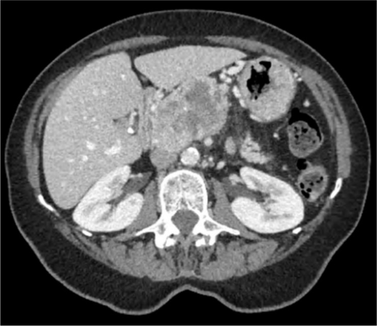
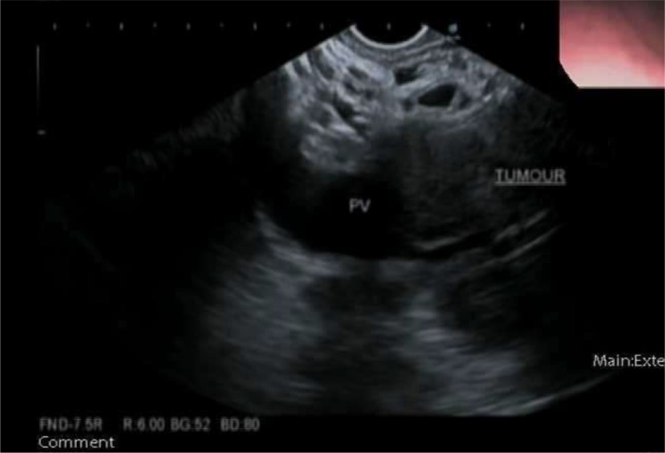
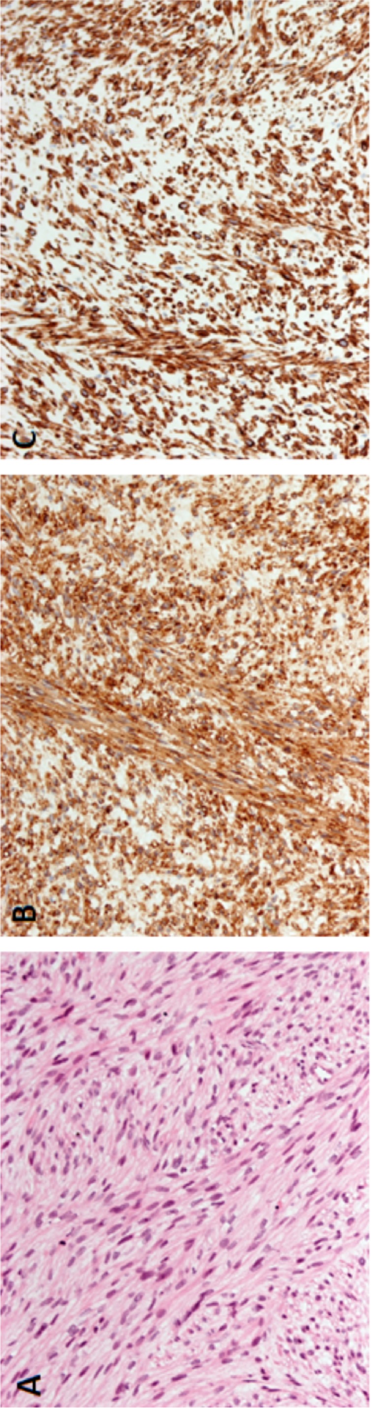
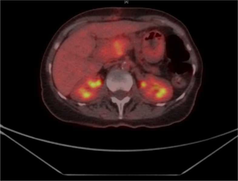
A 71-year-old woman was referred with abdominal pain and weight loss. An abdominal CT showed a 5-cm heterogeneous mass in the head of the pancreas with involvement of the superior mesenteric vein and artery. Her carcinoembryonic antigen (CEA) and CA 19-9 were normal. Two endoscopic ultrasound/fine needle aspirates (EUS/FNAs) of the mass diagnosed her with a mesenchymal tumour of myogenic origin but did not show features of malignancy. Frozen section analysis of laparoscopic core biopsies also failed to show malignant features, hence requiring an open biopsy which confirmed the diagnosis of pancreatic leiomyosarcoma (PLMS). She was eventually treated with radiotherapy. To our knowledge this is the only case in recent English literature of inoperable locally advanced PLMS that has required an open biopsy to formalise the diagnosis despite prior EUS FNAs. We include a review of the literature, highlighting the deficiencies of various biopsy techniques.
Keywords: general surgery; pancreas and biliary tract; pancreatic cancer; pathology.
© BMJ Publishing Group Limited 2019. No commercial re-use. See rights and permissions. Published by BMJ.
Conflict of interest statement
Competing interests: None declared.
Figures
References
-
- Brierley JD, Gospodarowicz MK, Wittekind C. Tnm classification of malignant tumours. John Wiley & Sons, 2016.
Publication types
MeSH terms
LinkOut - more resources
Full Text Sources
Medical