. 2019 Nov 12:13:508.
doi: 10.3389/fncel.2019.00508. eCollection 2019.

Synergy of Glutamatergic and Cholinergic Modulation Induces Plateau Potentials in Hippocampal OLM Interneurons

Affiliations

Synergy of Glutamatergic and Cholinergic Modulation Induces Plateau Potentials in Hippocampal OLM Interneurons

Nicholas Hagger-Vaughan et al. Front Cell Neurosci. .

Abstract

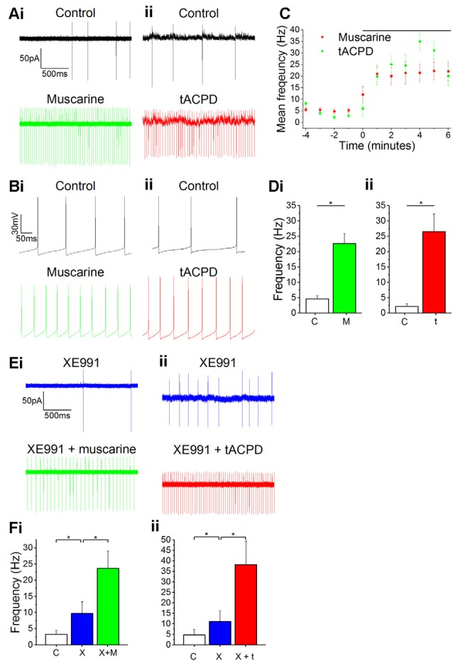
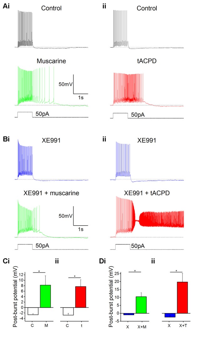
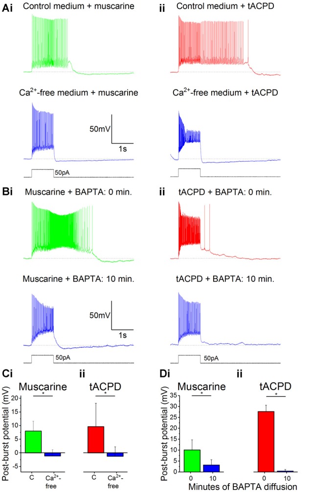
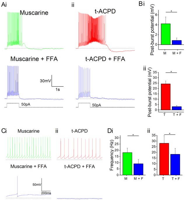
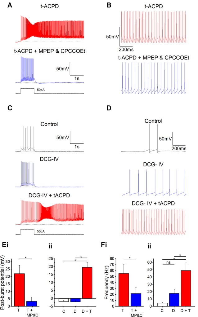
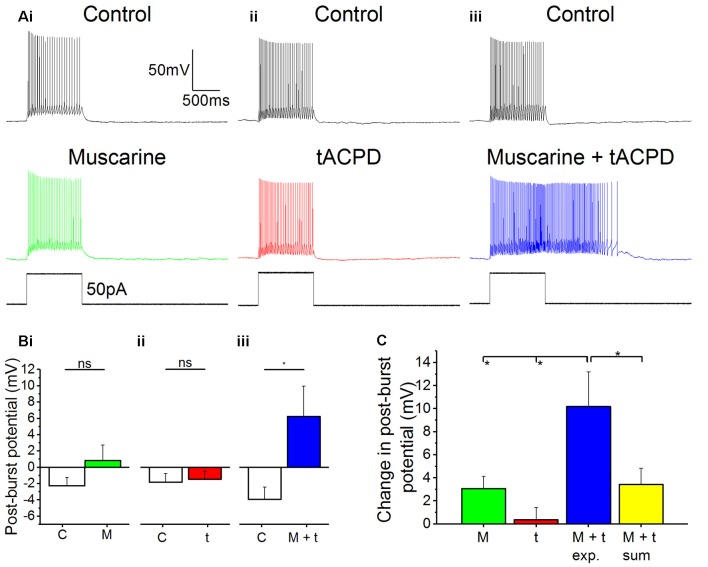
Oriens-lacunosum moleculare (OLM) cells are hippocampal inhibitory interneurons that are implicated in the regulation of information flow in the CA1 circuit, inhibiting cortical inputs to distal pyramidal cell dendrites, whilst disinhibiting CA3 inputs to pyramidal cells. OLM cells express metabotropic cholinergic (mAChR) and glutamatergic (mGluR) receptors, so modulation of these cells via these receptors may contribute to switching between functional modes of the hippocampus. Using a transgenic mouse line to identify OLM cells, we found that both mAChR and mGluR activation caused the cells to exhibit long-lasting depolarizing plateau potentials following evoked spike trains. Both mAChR- and mGluR-induced plateau potentials were eliminated by blocking transient receptor potential (TRP) channels, and were dependent on intracellular calcium concentration and calcium entry. Pharmacological tests indicated that Group I mGluRs are responsible for the glutamatergic induction of plateaus. There was also a pronounced synergy between the cholinergic and glutamatergic modulation, plateau potentials being generated by agonists applied together at concentrations too low to elicit any change when applied individually. This synergy could enable OLM cells to function as coincidence detectors of different neuromodulatory systems, leading to their enhanced and prolonged activation and a functional change in information flow within the hippocampus.

Keywords: OLM cells; TRP channel; interneuron; mAChR; mGluR; plateau potential; synergy.


Figures

Figure 1
Figure 1
Increased spontaneous firing rate following mAChR/mGluR activation is only partly mediated by M current. (A,B) Example traces showing spontaneous action potential firing observed during gap-free recording in control conditions in both cell-attached (A) and whole-cell (B) patch configurations. The frequency of firing increased in response to the application of either 10 μM muscarine (Ai,Bi) or 15 μM t-ACPD (Aii,Bii). (C) The rate of spontaneous firing during cell-attached recording in control conditions and the increase in frequency following the application of either muscarine or tACPD (presence of drug indicated by the black bar). Error bars = SEM. *p < 0.05. (D) The summary plot of the spontaneous firing frequency change for both muscarine (Di; n = 5) t-ACPD (Dii; n = 6). (E) Example traces showing an increase in spontaneous firing following the application of either muscarine (Ei) or tACPD (Eii) in addition to XE991. (F) The summary plot of the spontaneous firing frequency change following XE991 application, and application of either muscarine (Fi; n = 7) or t-ACPD (Fii; n = 6). Kv7 blockade increased the firing rate from the control and the application of mGluR or mAChR agonists significantly increased the firing rate further. Error bars = SEM. *P < 0.05.
Figure 2
Figure 2
mAChR and mGluR activation induce plateau potentials in oriens-lacunosum moleculare (OLM) cells. (A) Example traces showing the onset of a plateau potential following the application of either muscarine (Ai) or t-ACPD (Aii). (B) Example traces showing that blocking M-current with XE991 did not induce post-pulse spiking or plateau potentials, but these effects could be induced with additional application of muscarine (Bi) or tACPD (Bii). The horizontal, dotted lines in (A,B) indicate the baseline membrane potential during a 200 ms time window prior to the current pulse onset. (C) Summary plots showing the change in post-burst potential from control with either application of either muscarine (Ci; n = 6) or tACPD (Cii; n = 7), moving from an afterhyperpolarization (AHP) to an after-depolarization (ADP) in both conditions. (D) Summary plots showing the change in post-burst potential in the presence of XE 991 and following the application of either muscarine (Di; n = 6) or t-ACPD (Dii; n = 6). Both agonists induced a significant change in the post-pulse potential following in the presence of XE 991. Error bars = SEM. *P < 0.05.
Figure 3
Figure 3
The muscarinic and glutamatergic plateau potentials are calcium-dependent. (A) Example traces of the abolition of muscarine- (Ai) or tACPD-induced (Aii) plateau potentials by switching to a Ca2+-free artificial cerebrospinal fluid (aCSF) medium in the continuous presence of agonists. (B) Example traces showing the abolition of muscarine- (Bi) or tACPD-induced (Bii) plateau potentials immediately after (upper traces) and 10 min after break-in (lower traces) when the BAPTA-containing pipette solution became contiguous with the intracellular space. (C) Summary plots of the change in post-burst potential in the presence of either muscarine (Ci; n = 6) or tACPD (Cii; n = 7) after switching to a Ca2+-free medium, showing a move from an ADP to an AHP. (D) Summary plots of the change in post-burst potential in the presence of muscarine (Di; n = 5) or tACPD (Dii; n = 8) after 10 min of allowing BAPTA to diffuse into the intracellular solution. Error bars = SEM. *P < 0.05.
Figure 4
Figure 4
mAChR and mGluR agonist-induced effects can be reversed by a non-specific transient receptor potential (TRP) channel blocker, flufenamic acid (FFA). (A) Example traces showing plateau potentials induced (upper traces) by application of 10 μM muscarine (Ai) or 15 μM tACPD (Aii) are blocked by the addition of the non-specific TRP channel blocker, FFA (50 μM; lower traces). (B) Summary plots of post-pulse potential following application of FFA in the presence of either muscarine (Bi; n = 5) or tACPD (Bii; n = 4). In both cases, it was significantly reduced. (C) Example traces showing spontaneous firing in the presence of muscarine (Ci) or tACPD (Cii) before (upper trace) and after (lower trace) the addition of FFA. (D) Summary plots of the spontaneous firing rate following application of FFA in the presence of either muscarine (Di; n = 10) or tACPD (Dii; n = 10). Error bars = SEM. *P < 0.05. The firing rate significantly decreased in the presence of either mGluR or mAChR agonists.
Figure 5
Figure 5
Plateau potentials are evoked by group I but not group II mGluR activation. (A) Typical response to 50 pA 1 s current pulse in the presence of 15 μM tACPD, before and after application of the mGlu5R antagonist MPEP and the mGlu1R antagonist CPCCOEt. (B) Example traces showing the decrease in spontaneous firing in the presence of t-ACPD following addition of MPEP and CPCCOEt. (C) Example traces of plateau potentials following application of the selective Group II mGluR agonist DCG-IV and DCG-IV and tACPD. (D) Example traces of spontaneous firing following application of DCG-IV and DCG-IV and tACPD. (E) Summary plots of the post-pulse potential in the presence of t-ACPD following the addition of MPEP and CPCCOEt (i; n = 5) and following DCG-IV application and subsequence tACPD application (ii; n = 5). (F) Summary plots of spontaneous firing frequency in the presence of tACPD following the application of MPEP and CPCCOEt (i; n = 4), and in response to DCG-IV and following sequential application of tACPD (ii; n = 5). Error bars = SEM. *P < 0.05.
Figure 6
Figure 6
Synergistic effects on post-pulse potential by coapplication of low doses of muscarine and tACPD. (A) Typical responses of OLM cells to a 50 pA 1 s current pulse before and after application of either a low dose muscarine (Ai; 4 μM; n = 8), a low dose tACPD (Aii; 4 μM; n = 8), or both combined (Aiii; 4 μM + 4 μM; n = 6). (B) Summary plots show that no significant change in post-burst potential was seen with application of a low dose of muscarine (Bi; n = 8) or tACPD (Bii; n = 8), whereas a significant increase was seen following application of a low doses of both drugs simultaneously (Biii; n = 6), converting the post-burst potential from and AHP to ADP. (C) The Summary plot for change in post-pulse potential, in all experimental conditions and also post hoc linear sum (yellow column). The linear sum was calculated from eight randomly selected pairs of independent measures of single low dose drug administration. Error bars = SEM. *P < 0.05; ns, not significantly different, P > 0.05.

Similar articles

Cited by

References

    1. Azouz R., Jensen M. S., Yaari Y. (1994). Muscarinic modulation of intrinsic burst firing in rat hippocampal neurons. Eur. J. Neurosci. 6, 961–966. 10.1111/j.1460-9568.1994.tb00590.x - DOI - PubMed
    1. Ben-Mabrouk F., Amos L. B., Tryba A. K. (2012). Metabotropic glutamate receptors (mGluR5) activate TRPC channels to improve the regularity of the respiratory rhythm generated by the pre-Bötzinger Complex in mice. Eur. J. Neurosci. 35, 1725–1737. 10.1111/j.1460-9568.2012.08091.x - DOI - PMC - PubMed
    1. Bittner K. C., Milstein A. D., Grienberger C., Romani S., Magee J. C. (2017). Behavioral time scale synaptic plasticity underlies CA1 place fields. Science 357, 1033–1036. 10.1126/science.aan3846 - DOI - PMC - PubMed
    1. Blasco-Ibáñez J. M., Freund T. F. (1995). Synaptic input of horizontal interneurons in stratum oriens of the hippocampal CA1 subfield: structural basis of feed-back activation. Eur. J. Neurosci. 7, 2170–2180. 10.1111/j.1460-9568.1995.tb00638.x - DOI - PubMed
    1. Bocchio M., Lukacs I. P., Stacey R., Plaha P., Apostolopoulos V., Livermore L., et al. . (2019). Group II metabotropic glutamate receptors mediate presynaptic inhibition of excitatory transmission in pyramidal neurons of the human cerebral cortex. Front. Cell. Neurosci. 12:508. 10.3389/fncel.2018.00508 - DOI - PMC - PubMed

LinkOut - more resources