

Complete Multilineage CD4 Expression Defect Associated With Warts Due to an Inherited Homozygous CD4 Gene Mutation
- PMID: 31781092
- PMCID: PMC6856949
- DOI: 10.3389/fimmu.2019.02502
Complete Multilineage CD4 Expression Defect Associated With Warts Due to an Inherited Homozygous CD4 Gene Mutation
Abstract
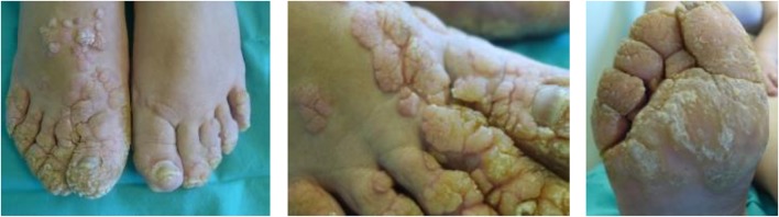
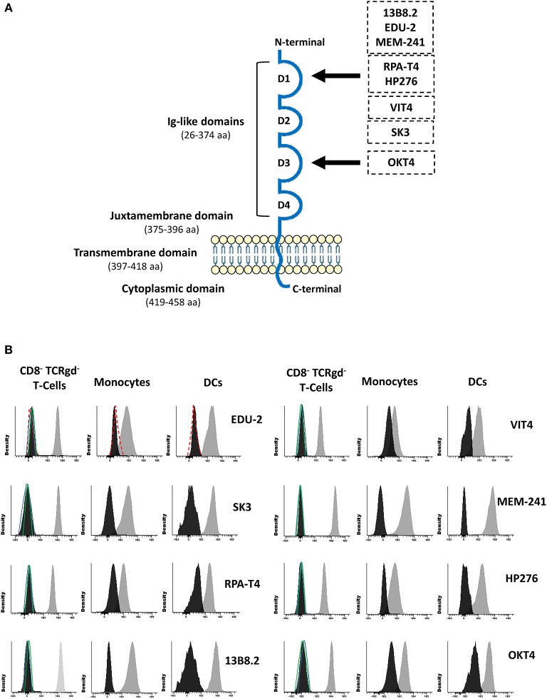
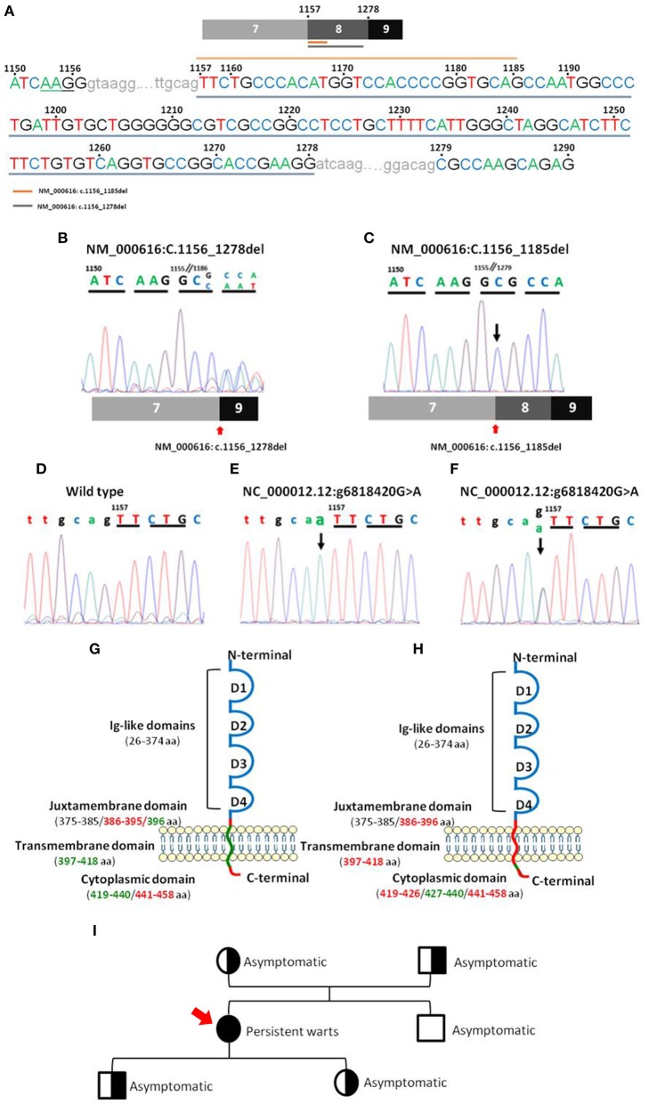
Idiopathic T-CD4 lymphocytopenia (ICL) is a rare and heterogeneous syndrome characterized by opportunistic infections due to reduced CD4 T-lymphocytes (<300 cells/μl or <20% T-cells) in the absence of HIV infection and other primary causes of lymphopenia. Molecular testing of ICL has revealed defects in genes not specific to CD4 T-cells, with pleiotropic effects on other cell types. Here we report for the first time an absolute CD4 lymphocytopenia (<0.01 CD4+ T-cells/μl) due to an autosomal recessive CD4 gene mutation that completely abrogates CD4 protein expression on the surface membrane of T-cells, monocytes, and dendritic cells. A 45-year-old female born to consanguineous parents consulted because of exuberant, relapsing, and treatment-refractory warts on her hands and feet since the age of 10 years, in the absence of other recurrent infections or symptoms. Serological studies were negative for severe infections, including HIV 1/2, HTLV-1, and syphilis, but positive for CMV and EBV. Blood analysis showed the absence of CD4+ T-cells (<0.01%) with repeatedly increased counts of B-cells, naïve CD8+ T-lymphocytes, and particularly, CD4/CD8 double-negative (DN) TCRαβ+ TCRγδ- T-cells (30% of T-cells; 400 cells/μl). Flow cytometric staining of CD4 using monoclonal antibodies directed against five different epitopes, located in two different domains of the protein, confirmed no cell surface membrane or intracytoplasmic expression of CD4 on T-cells, monocytes, and dendritic cells but normal soluble CD4 plasma levels. DN T-cells showed a phenotypic and functional profile similar to normal CD4+ T-cells as regards expression of maturation markers, T-helper and T-regulatory chemokine receptors, TCRvβ repertoire, and in vitro cytokine production against polyclonal and antigen-specific stimuli. Sequencing of the CD4 gene revealed a homozygous (splicing) mutation affecting the last bp on intron 7-8, leading to deletion of the juxtamembrane and intracellular domains of the protein and complete abrogation of CD4 expression on the cell membrane. These findings support previous studies in CD4 KO mice suggesting that surrogate DN helper and regulatory T-cells capable of supporting antigen-specific immune responses are produced in the absence of CD4 signaling and point out the need for better understanding the role of CD4 on thymic selection and the immune response.
Keywords: CD4; CD4 lymphopenia; double-negative T-cells (DNTs); idiopathic CD4 lymphocytopenia; warts.
Copyright © 2019 Fernandes, Perez-Andres, Blanco, Jara-Acevedo, Criado, Almeida, Botafogo, Coutinho, Paiva, van Dongen, Orfao and Faria.
Figures
Similar articles
-
Humanized mouse models reveal an immunologic classification of idiopathic CD4 lymphocytopenia subtypes.JCI Insight. 2019 Jul 25;4(14):e127802. doi: 10.1172/jci.insight.127802. eCollection 2019 Jul 25. JCI Insight. 2019. PMID: 31341106 Free PMC article.
-
Primary T-cell immunodeficiency with immunodysregulation caused by autosomal recessive LCK deficiency.J Allergy Clin Immunol. 2012 Nov;130(5):1144-1152.e11. doi: 10.1016/j.jaci.2012.07.029. Epub 2012 Sep 15. J Allergy Clin Immunol. 2012. PMID: 22985903
-
Idiopathic CD4+ T lymphopenia without autoimmunity or granulomatous disease in the slipstream of RAG mutations.Blood. 2011 Jun 2;117(22):5892-6. doi: 10.1182/blood-2011-01-329052. Epub 2011 Apr 18. Blood. 2011. PMID: 21502542
-
Idiopathic lymphocytopenia.Curr Opin Hematol. 2015 Jan;22(1):46-52. doi: 10.1097/MOH.0000000000000102. Curr Opin Hematol. 2015. PMID: 25463685 Review.
-
Idiopathic CD4 lymphocytopenia: a case of missing, wandering or ineffective T cells.Arthritis Res Ther. 2012 Aug 31;14(4):222. doi: 10.1186/ar4027. Arthritis Res Ther. 2012. PMID: 22971990 Free PMC article. Review.
Cited by
-
The monogenic landscape of human infectious diseases.J Allergy Clin Immunol. 2025 Mar;155(3):768-783. doi: 10.1016/j.jaci.2024.12.1078. Epub 2024 Dec 24. J Allergy Clin Immunol. 2025. PMID: 39724971 Review.
-
Human genetic dissection of papillomavirus-driven diseases: new insight into their pathogenesis.Hum Genet. 2020 Jun;139(6-7):919-939. doi: 10.1007/s00439-020-02183-x. Epub 2020 May 20. Hum Genet. 2020. PMID: 32435828 Free PMC article. Review.
-
Human genetic and immunological dissection of papillomavirus-driven diseases: new insights into their pathogenesis.Curr Opin Virol. 2021 Dec;51:9-15. doi: 10.1016/j.coviro.2021.09.002. Epub 2021 Sep 21. Curr Opin Virol. 2021. PMID: 34555675 Free PMC article. Review.
-
Infections in the monogenic autoimmune syndrome APECED.Curr Opin Immunol. 2021 Oct;72:286-297. doi: 10.1016/j.coi.2021.07.011. Epub 2021 Aug 18. Curr Opin Immunol. 2021. PMID: 34418591 Free PMC article. Review.
-
In-depth blood immune profiling of Good syndrome patients.Front Immunol. 2023 Nov 15;14:1285088. doi: 10.3389/fimmu.2023.1285088. eCollection 2023. Front Immunol. 2023. PMID: 38035080 Free PMC article.
References
Publication types
MeSH terms
Substances
LinkOut - more resources
Full Text Sources
Medical
Molecular Biology Databases
Research Materials