. 2019 Nov 8:10:1401.
doi: 10.3389/fpls.2019.01401. eCollection 2019.

Membrane-Specific Targeting of Tail-Anchored Proteins SECE1 and SECE2 Within Chloroplasts

Affiliations

Membrane-Specific Targeting of Tail-Anchored Proteins SECE1 and SECE2 Within Chloroplasts

Stacy A Anderson et al. Front Plant Sci. .

Abstract

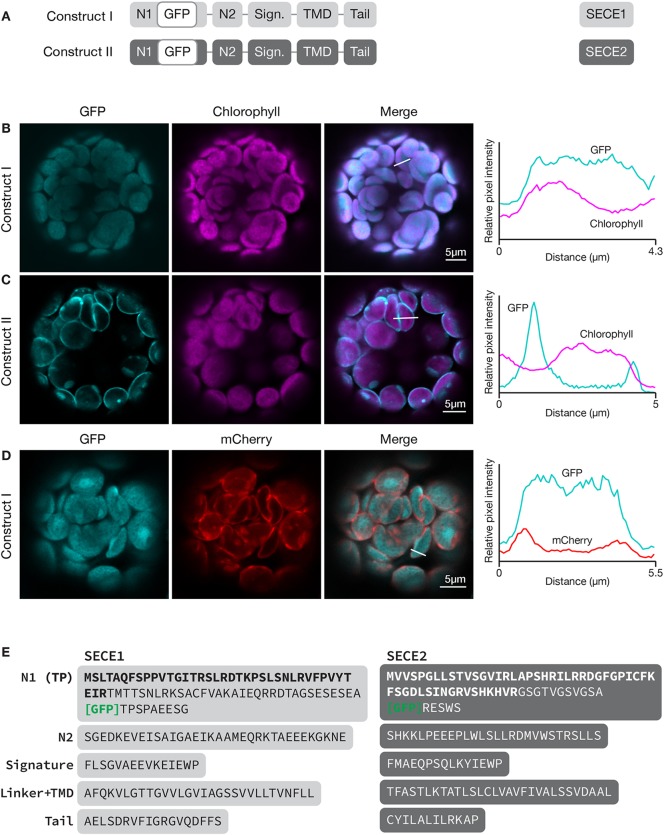
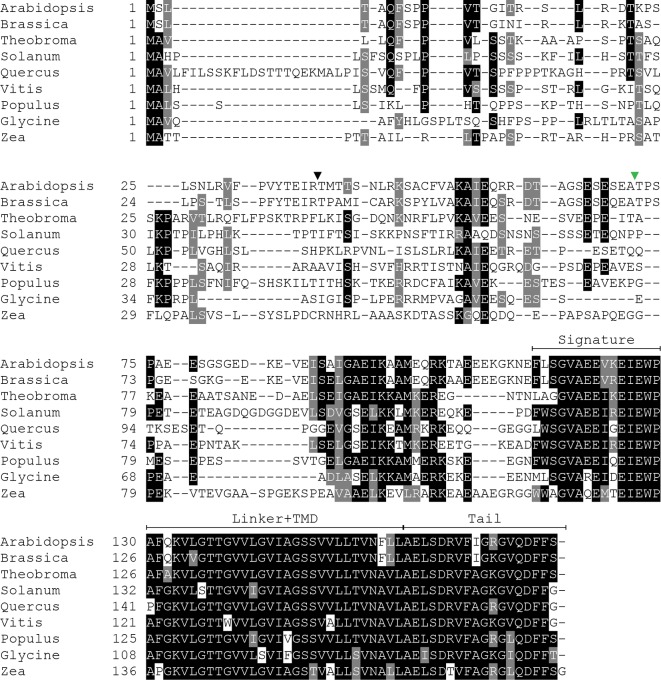
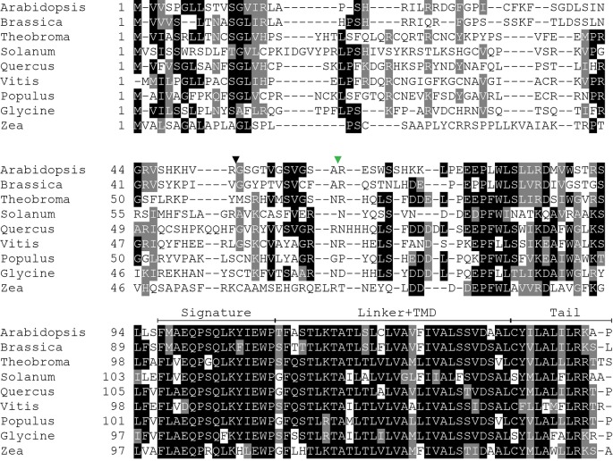
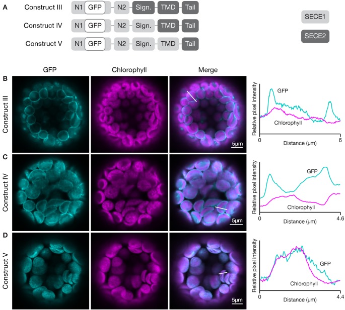
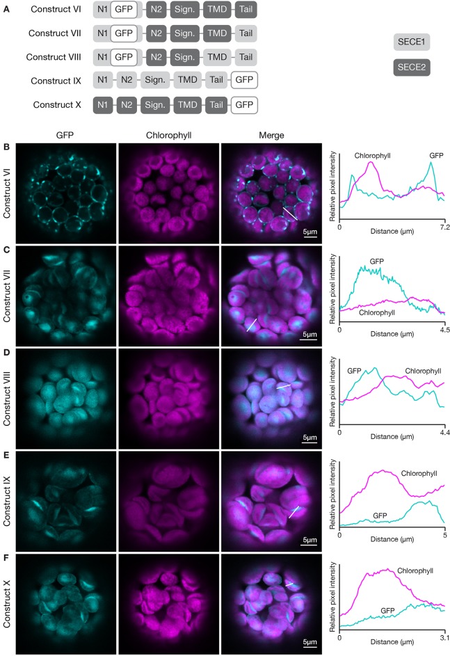
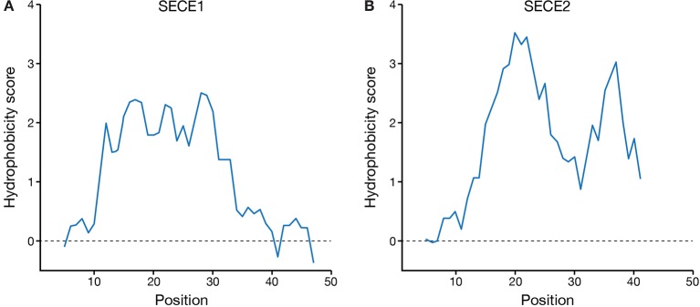
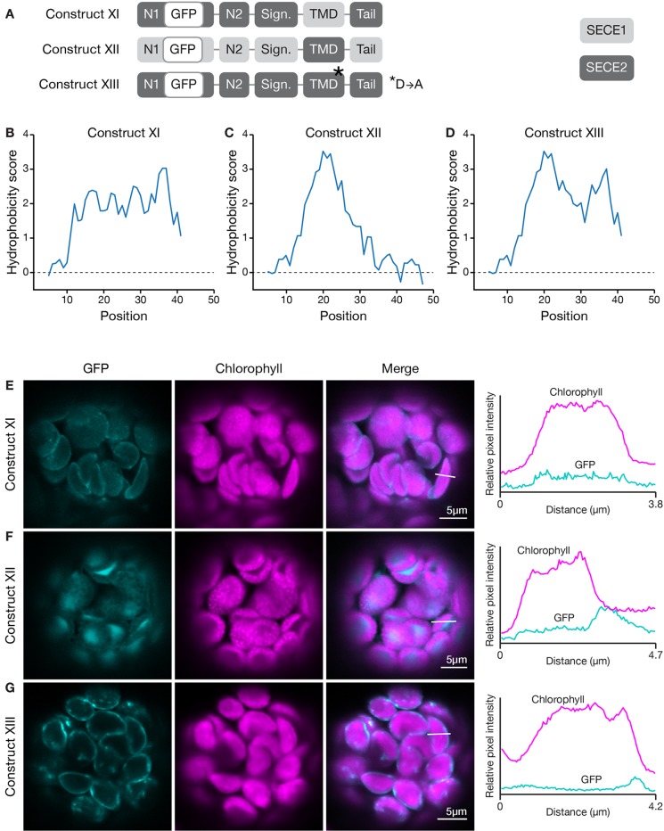
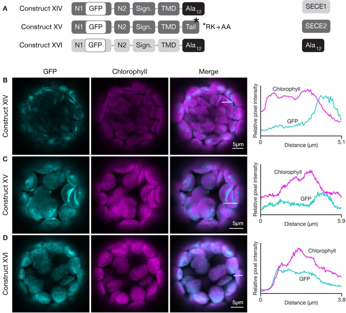
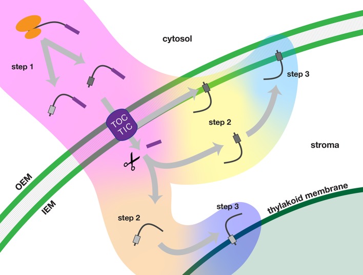
Membrane proteins that are imported into chloroplasts must be accurately targeted in order to maintain the identity and function of the highly differentiated internal membranes. Relatively little is known about the targeting information or pathways that direct proteins with transmembrane domains to either the inner envelope or thylakoids. In this study, we focused on a structurally simple class of membrane proteins, the tail-anchored proteins, which have stroma-exposed amino-terminal domains and a single transmembrane domain within 30 amino acids of the carboxy-terminus. SECE1 and SECE2 are essential tail-anchored proteins that function as components of the dual SEC translocases in chloroplasts. SECE1 localizes to the thylakoids, while SECE2 localizes to the inner envelope. We have used transient expression in Arabidopsis leaf protoplasts and confocal microscopy in combination with a domain-swapping strategy to identify regions that contain important targeting determinants. We show that membrane-specific targeting depends on features of the transmembrane domains and the short C-terminal tails. We probed the contributions of these regions to targeting processes further through site-directed mutagenesis. We show that thylakoid targeting still occurs when changes are made to the tail of SECE1, but changing residues in the tail of SECE2 abolishes inner envelope targeting. Finally, we discuss possible parallels between sorting of tail-anchored proteins in the stroma and in the cytosol.

Keywords: SEC translocase; chloroplast; inner envelope membrane; organelle biogenesis; tail-anchored protein; targeting; thylakoid.


Figures

Figure 1
Figure 1
Localization of GFP-SECE1 and GFP-SECE2 in transfected protoplasts and designation of protein domains in SECE1 and SECE2. (A) Diagrams depicting constructs I and II. SECE1 sequences are indicated with light gray and SECE2 sequences with dark gray; N1 and N2: N-terminal regions; GFP: green fluorescent protein; Sign.: signature domain; TMD: linker plus transmembrane domain; Tail: C-terminal tail. (BD) Leaf protoplasts from 4–5 week old wildtype (Columbia ecotype) plants were transfected with (B) construct I, (C) construct II, or (D) construct I plus a TIC20-mCherry construct. The images show GFP fluorescence (cyan), chlorophyll fluorescence (magenta) or TIC20-mCherry fluorescence (red), merged images, and relative pixel intensity diagrams that correspond with the white lines on the merged images. (E) Amino acid sequences that correspond to the individual domains of SECE1 and SECE2. Predicted transit peptides (TP) are shown in bold text and the GFP insertion site is indicated. TMD, transmembrane domain.
Figure 2
Figure 2
Multiple sequence alignment of SECE1 from a variety of plant species. The predicted transit peptide cleavage site in Arabidopsis is indicated with a black triangle. The GFP insertion site is indicated by a green triangle. Signature, signature region; TMD, transmembrane domain; Tail, C-terminal tail.
Figure 3
Figure 3
Multiple sequence alignment of SECE2 from a variety of plant species. The predicted transit peptide cleavage site in Arabidopsis is indicated with a black triangle. The GFP insertion site is indicated by a green triangle. Signature, signature region; TMD, transmembrane domain; Tail, C-terminal tail.
Figure 4
Figure 4
Localization of fluorescent chimeric proteins containing N-terminal SECE1 regions and C-terminal SECE2 regions in transfected protoplasts. (A) Diagrams depicting constructs III, IV, and V. SECE1 sequences are indicated with light gray and SECE2 sequences with dark gray; N1 and N2: N-terminal region; GFP: green fluorescent protein; Sign.: signature domain; TMD: linker plus transmembrane domain; Tail: C-terminal tail. (BD) Leaf protoplasts from 4–5 week old wildtype (Columbia ecotype) plants were transfected with (B) construct III, (C) construct IV, or (D) construct V. The images show GFP fluorescence (cyan), chlorophyll fluorescence (magenta), merged images, and relative pixel intensity diagrams that correspond with the white lines on the merged images.
Figure 5
Figure 5
Localization of fluorescent chimeric SECE1 and SECE2 proteins in transfected protoplasts. The SECE1 N1-region is included in constructs VI–VIII to facilitate chloroplast import. (A) Diagrams depicting constructs VI, VII, VIII, IX, and X. SECE1 sequences are indicated with light gray and SECE2 sequences with dark gray; N1 and N2: N-terminal region; GFP: green fluorescent protein; Sign.: signature domain; TMD: linker plus transmembrane domain; Tail: C-terminal tail. (BF) Leaf protoplasts from 4–5 week old wildtype (Columbia ecotype) plants were transfected with (B) construct VI, (C) construct VII, (D) construct VIII, (E) construct IX, or (F) construct X. The images show GFP fluorescence (cyan), chlorophyll fluorescence (magenta), merged images, and relative pixel intensity diagrams that correspond with the white lines on the merged images.
Figure 6
Figure 6
Hydrophobicity profiles of the C-terminal regions of SECE1 and SECE2. Profiles were generated through ExPASy ProtScale (https://web.expasy.org/protscale/) using the Kyte & Doolittle amino acid scale with a window size of 9. Each profile represents the linker, TMD, and tail regions beginning with the E-W-P motif. (A) Hydrophobicity profile of SECE1’s C-terminal regions, corresponding to residues 127–177 of the full-length protein. (B) Hydrophobicity profile of SECE2’s C-terminal regions, corresponding to residues 109–153 of the full-length protein.
Figure 7
Figure 7
Localization of fluorescent chimeric SECE1 and SECE2 proteins with alterations in the TMD region. (A) Diagrams depicting constructs XI, XII, and XIII. SECE1 sequences are indicated with light gray and SECE2 sequences with dark gray; N1 and N2: N-terminal regions; GFP: green fluorescent protein; Sign.: signature domain; TMD: linker plus transmembrane domain; Tail: C-terminal tail. The asterisk indicates the location of a D to A amino acid substitution. (BD) Hydrophobicity profiles of the linker, TMD, and tail regions of the proteins encoded by (B) construct XI, (C) construct XII, and (D) construct XIII. (E–G) Leaf protoplasts from 4–5 week old wildtype (Columbia ecotype) plants were transfected with (E) construct XI, (F) construct XII, or (G) construct XIII. The images show GFP fluorescence (cyan), chlorophyll fluorescence (magenta), merged images, and relative pixel intensity diagrams that correspond with the white lines on the merged images.
Figure 8
Figure 8
Localization of fluorescent chimeric SECE1 and SECE2 proteins with alterations in the C-terminal tail regions. (A) Diagrams depicting constructs XIV, XV, and XVI. SECE1 sequences are indicated with light gray and SECE2 sequences with dark gray; N1 and N2: N-terminal regions; GFP: green fluorescent protein; Sign.: signature domain; TMD: linker plus transmembrane domain; Tail: C-terminal tail. The black rectangles with Ala12 indicate a substitution of the entire native tail with 12 alanine residues. The asterisk indicates the location of an R-K to A-A amino acid substitution. (B–D) Leaf protoplasts from 4–5 week old wildtype (Columbia ecotype) plants were transfected with (B) construct XIV, (C) construct XV, or (D) construct XVI. The images show GFP fluorescence (cyan), chlorophyll fluorescence (magenta), merged images, and relative pixel intensity diagrams that correspond with the white lines on the merged images.
Figure 9
Figure 9
Multi-step model of targeting nuclear-encoded TA proteins to the inner envelope or thylakoid membranes of chloroplasts. In step 1 (pink background color), the TA protein is synthesized in the cytosol and delivered to the chloroplast. In step 2, the TA proteins are sorted either in an inner envelope-directed (yellow background color) or thylakoid-directed (peach background color) pathway. In step 3, the TA proteins are integrated either into the inner envelope membrane (light blue background color) or the thylakoid membrane (dark blue background). Scissors indicate stromal processing peptidase activity. IEM, inner envelope membrane; OEM, outer envelope membrane; TOC/TIC, Translocons of the Outer and Inner Envelope membranes of the chloroplast.

Similar articles

Cited by

References

    1. Altschul S. F., Gish W., Miller W., Myers E. W., Lipman D. J. (1990). Basic local alignment search tool. J. Mol. Biol. 3, 403–410. 10.1016/S0022-2836(05)80360-2 - DOI - PubMed
    1. Benning C. (2009). Mechanisms of lipid transport involved in organelle biogenesis in plant cells. Annu. Rev. Cell Dev. Biol. 25, 71–91. 10.1146/annurev.cellbio.042308.113414 - DOI - PubMed
    1. Bionda T., Tillmann B., Simm S., Beilstein K., Ruprecht M., Schleiff E. (2010). Chloroplast import signals: The length requirement for translocation in vitro and in vivo. J. Mol. Biol. 402, 510–523. 10.1016/j.jmb.2010.07.052 - DOI - PubMed
    1. Bölter B., Soll J. (2016). Once upon a time – chloroplast protein import research from infancy to future challenges. Mol. Plant 9, 798–812. 10.1016/j.molp.2016.04.014 - DOI - PubMed
    1. Chio U. S., Cho H., Shan S. (2017). Mechanisms of tail-anchored membrane protein targeting and insertion. Annu. Rev. Cell Dev. Biol. 33, 417–438. 10.1146/annurev-cellbio-100616-060839 - DOI - PMC - PubMed

LinkOut - more resources