Review
. 2019 Nov 12:10:1435.
doi: 10.3389/fpls.2019.01435. eCollection 2019.

Development of Parasitic Organs of a Stem Holoparasitic Plant in Genus Cuscuta

Affiliations
Review

Development of Parasitic Organs of a Stem Holoparasitic Plant in Genus Cuscuta

Kohki Shimizu et al. Front Plant Sci. .

Abstract

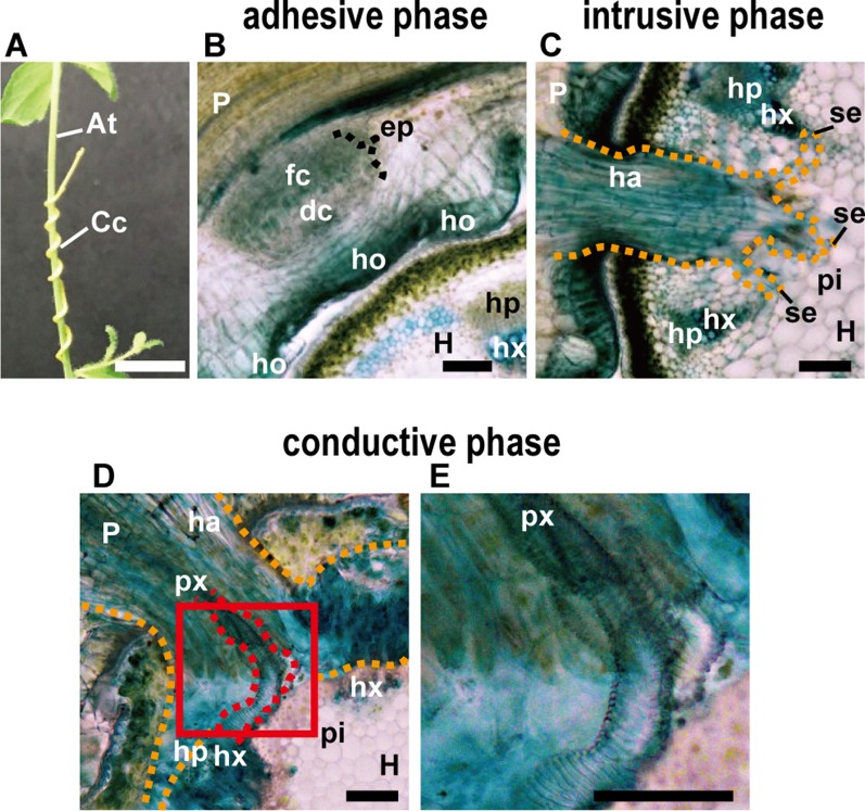
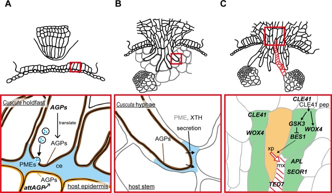
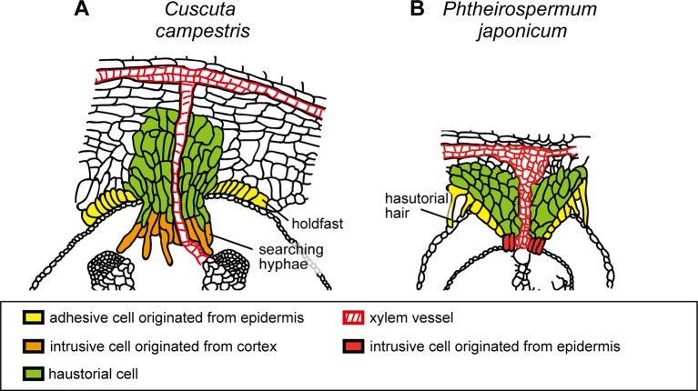
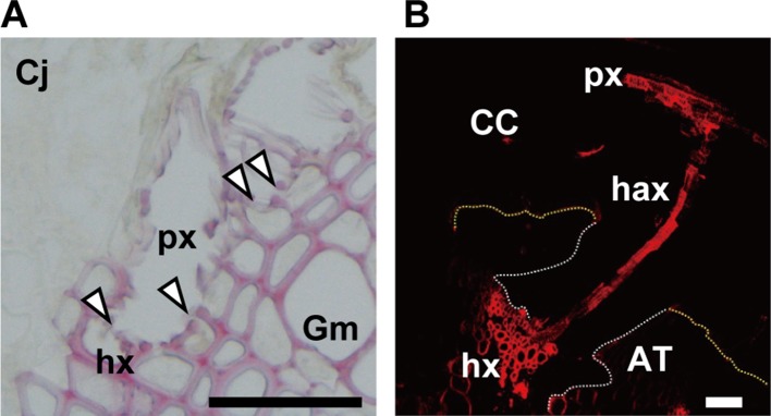
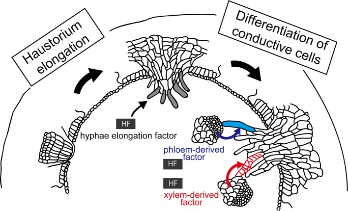
Parasitic plants infect a broad range of plant species including economically important crops. They survive by absorbing water, minerals, and photosynthates from their hosts. To support their way of life, parasitic plants generally establish parasitic organs that allow them to attach to their hosts and to efficiently absorb substances from the vascular system of the host. Here, we summarize the recent progress in understanding the mechanisms underlying the formation of these parasitic organs, focusing on the process depicted in the stem holoparasitic genus, Cuscuta. An attachment structure called "holdfast" on the stem surface is induced by the light and contact stimuli. Concomitantly with holdfast formation, development of an intrusive structure called haustorium initiates in the inner cortex of the Cuscuta stem, and it elongates through apoplastic space of the host tissue. When haustoria reaches to host vascular tissues, they begin to form vascular conductive elements to connect vascular tissue of Cuscuta stem to those of host. Recent studies have shown parasite-host interaction in the interfacial cell wall, and regulation of development of these parasitic structures in molecular level. We also briefly summarize the role of host receptor in the control of compatibility between Cuscuta and hosts, on which occurrence of attachment structure depends, and the role of plant-to-plant transfer of long-distance signals after the establishment of conductive structure.

Keywords: Cuscuta; attachment cells; conductive cells; haustorium; host factors; intrusive cells; parasitic organs; parasitic plants.


Figures

Figure 1
Figure 1
(A) Appearance of parasitic site formed between Cuscuta campestris (Cc) and Arabidopsis thaliana (At) from the outside. C. campestris coils around the inflorescence stem of Arabidopsis. Scale bar, 1 cm. (B–E) Transverse sections of the three phases of parasitic processes of Cuscuta. Scale bars, 200 μm. (B) Adhesive phase. Holdfast (ho) is formed on the host-attaching surface of C. campestris. Prehaustorium develops in the inner cortex of the stem right behind holdfast. In the endophyte primordium (ep), digitate cells (dc) and file cells (fc) differentiate and start to elongate. (C) Intrusive phase. Haustorium (ha) intrudes in the cortex of the host stem. It sometimes reaches to the pith (pi). (D) Conductive phase. (E) Area in the red square in (D) is magnified. Vascular conductive elements (px) are formed in the haustorim. P, parasite; H, host; ha, haustorium; hp, host phloem; hx, host xylem; px, parasite xylem; pi, pith; se, searching hypha; orange dotted line, outline of haustorium; red dotted line, outline of parasite xylem. In all panels, 200-μm-thick micro-slicer sections were stained with toluidine blue.
Figure 2
Figure 2
Functions of enzymes and genes associated with the parasitic processes. Panels in the bottom show magnified views of the areas in red squares in panels on the top. (A) Putative function of cell wall-modifying enzymes secreted from holdfast in the adhesive phase. Holdfast cells tighten the adhesion by pectin-rich cement (ce, blue). It has been shown that holdfast cells of Cuscuta campestris contain numerous secretion vesicles containing the components of cell-wall-loosening complexes. Pectin methylesterases (PMEs) are probably secreted to tighten the adhesion of Cuscuta to host. Specific members of genes encoding arabinogalactan proteins (AGPs) are expressed in searching hyphae, and accumulate AGP proteins (brown). AGP also have roles in host cell surface (orange) in the adhesion of parasite (Albert et al., 2006). (B) Secretion of cell wall-modifying enzymes to the cell walls adjacent to searching hyphae in the intrusive phase. Xyloglucan endotransglucosylation (XET) activity of XTH was detected in interface (blue) at the tip of haustoria of C. reflexa (Olsen and Krause, 2017). In C. campestris, searching hyphae-specific expression of FASCICLIN-LIKE genes causes the accumulation of AGPs in the interfacial cell walls surrounding searching hypha cells (brown) (Hozumi et al., 2017) Exact role of AGPs in the intrusive phase is still unknown. (C) Expression of genes associated with differentiation of vascular elements during the transition from intrusive phase to conductive phase in haustorim of Cuscuta japonica. Green, procambium/phloem region, orange, xylem precursor (xp), red diagonal lines, mature xylem vessel (mx). WOX4, WUSCHEL RELATED HOMEOBOX 4; CLE41, CLAVATA3/EMBRYO SURROUNDING REGION-RELATED 41; GSK3, GLYCOGEN SYNTHASE KINASE 3; BES1, BRI1-EMS-SUPPRESSOR 1; TED7, TRACHEARY ELEMENT DIFFERENTIATION-RELATED 7; APL, ALTERED PHLOEM DEVELOPMENT; SEOR1, SIEVE ELEMENT OCCLUSION-RELATED 1.
Figure 3
Figure 3
Schematic illustration of the structures of haustoria of Cuscuta campestris and Phtheirospermum japonicum. (A) Cuscuta campestris, a holoparasitic plant belonging to Convolvulaceae, develops lateral haustoria. (B) Phtheirospermum japonicum, a hemiparasitic plant belonging to Orobanchaceae, develops lateral haustoria. Holdfast of C. campestris and haustorial hair of P. japonicum are likely to be analogous that develop from epidermal cells and contribute to the adhesion of parasite to host. Intrusive cells of C. campestris, searching hyphae, develop from digitate cells which have been differentiated from the cortex or endodermal cells of the stem. On the other hand, intrusive cells of P. japonicum are shown to be differentiated from the epidermal cells.
Figure 4
Figure 4
(A) Open connection (arrowheads) between xylem vessels of parasite (px) and host (hx) in the parasitic interface of Cuscuta japonica (Cj) with Glycine max (Gm). Scale bar, 50 μm. A 20-μm-thick paraffin-embedded section was stained with phloroglucinol. (B) Transfer of 5-carboxytetramethylrhodamine (TMR) 10-kDa dextran (red) from host xylem vessel to haustorial xylem vessels, and then to Cuscuta stem xylem. White dotted line; outline of haustorium, yellow dotted line; outline of attachment boundary between Cuscuta and host Arabidopsis. CC, Cuscuta campestris; AT, Arabidopsis thaliana; px, parasite xylem vessel; hax, haustorial xylem vessel; hx, host xylem vessel. Scale bar, 200 μm.
Figure 5
Figure 5
Involvement of host factors (HFs) in the elongation and differentiation of searching hyphae. (Left) HF inducing elongation of searching hyphae (left) has been hypothesized because digitate cells or file cells of C. campestris initiated in a host-independent manner do not show further development of searching hyphae without host. (Right) HF has been implied in the differentiation of searching hypha cells into xylem (red lines) and phloem conductive elements (blue), because upon contacting xylem vessels or phloem sieve tubes of the host, the hyphal cells starts to differentiate into respective conductive elements. These HFs have not been identified yet.

Similar articles

Cited by

References

    1. Alakonya A., Kumar R., Koenig D., Kimura S., Townsley B., Runo S., et al. , (2012). Interspecific RNA interference of SHOOT MERISTEMLESS-like disrupts Cuscuta pentagona plant parasitism. Plant Cell 24, 3153–3166. 10.1105/tpc.112.099994 - DOI - PMC - PubMed
    1. Albert M., Werner M., Proksch P., Fry S. C., Kaldenhoff R. (2004). The cell wall-modifying xyloglucan endotransglycosylase/hydrolase LeXTH1 is expressed during the defence reaction of tomato against the plant parasite Cuscuta reflexa. Plant Biol. 6, 402–407. 10.1055/s-2004-817959 - DOI - PubMed
    1. Albert M., Belastegui-Macadam X., Kaldenhoff R. (2006). An attack of the plant parasite Cuscuta reflexa induces the expression of attAGP, an attachment protein of the host tomato. Plant J. 48, 548–556. 10.1111/j.1365-313X.2006.02897.x - DOI - PubMed
    1. Birschwilks M., Haupt S., Hofius D., Neumann S. (2006). Transfer of phloem-mobile substances from the host plants to the holoparasite Cuscuta sp. J. Exp. Bot. 57, 911–921. 10.1093/jxb/erj076 - DOI - PubMed
    1. Bleischwitz M., Albert M., Fuchsbauer H. L., Kaldenhoff R. (2010). Significance of Cuscutain, a cysteine protease from Cuscuta reflexa, in host-parasite interactions. BMC Plant Biol. 10, 227. 10.1186/1471-2229-10-227 - DOI - PMC - PubMed

LinkOut - more resources