. 2019 Oct 30:2019:3091619.
doi: 10.1155/2019/3091619. eCollection 2019.

Synergistic Effects of Human Platelet-Rich Plasma Combined with Adipose-Derived Stem Cells on Healing in a Mouse Pressure Injury Model

Affiliations

Synergistic Effects of Human Platelet-Rich Plasma Combined with Adipose-Derived Stem Cells on Healing in a Mouse Pressure Injury Model

Zhiyuan Liu et al. Stem Cells Int. .

Abstract

Pressure injury (PI) affects quality of life and results in economic and social burdens. Local transplantation of human adipose-derived stem cells (ASCs) is considered an effective treatment. However, ASC suspension alone is vulnerable to the immune system and results in a shortened cell survival. There is increasing evidence of a synergistic effect of platelet-rich plasma (PRP) combined with ASCs on wound healing. This study investigated the effectiveness, synergy, and mechanism of wound healing following local injection of PRP combined with ASCs in a rodent PI model. PRP or ASCs alone were the control intervention. Wound healing, inflammatory infiltration, collagen deposition, angiogenesis, neurogenesis, and cell homing were investigated. PI healing was promoted by the synergistic effects of PRP combined with ASCs. The combination was more effective than ASCs alone for modulating inflammation, increasing collagen deposition, angiogenesis, neurogenesis, and the persistence of the injected ASCs. These data provide a theoretical foundation for the clinical administration of ASCs combined with PRP in PI healing and skin regeneration.


Conflict of interest statement

The authors have no competing financial interests to declare.

Figures

Figure 1
Figure 1
ASCs from human subcutaneous adipose tissue had characteristics of mesenchymal stem cells. Representative phase-contrast micrographs of ASCs show fibroblast morphology. The percentage of GFP-transfected cells was high; the morphology did not change significantly (a). ASCs differentiated into adipocyte-, osteoblast-, and cartilage-like cells (×100 magnification) (b). Flow cytometry of ASCs (c). ASCs: adipose-derived stem cells.
Figure 2
Figure 2
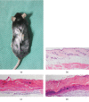
Animal model of pressure injury (PI) (a). Normal skin structure (b). On day 1 after the establishment of the PI model, skin thinning, loss of the normal skin structure, and inflammatory cell infiltration (∗) are visible (c). On day 5, the epidermis was exfoliated, degeneration and necrosis of subcutaneous tissue are visible, and infiltration of inflammatory cells is extensive (∗) (d). ×100 magnification.
Figure 3
Figure 3
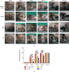
PRP combined with ASCs, ASCs, or PRP alone was injected subdermally at the wound margins. Healing of pressure wounds was monitored. Representative images of wound healing at increasing times after local transplantation of PRP combined with ASCs, ASCs, or PRP (a). Histogram of wound healing over 17 days showing significant differences in the rate of healing (b). ∗∗P < 0.01, ∗∗∗P < 0.001. ASCs: human adipose-derived stem cells; PRP: platelet-rich plasma.
Figure 4
Figure 4
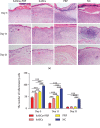
PRP combined with ASCs, ASCs, or PRP alone suppressed the inflammatory response to wounding. Tissue histology shows that inflammatory cell infiltration in the dermis and subcutaneous layers on days 5 and 11 was less in tissue from ASC plus PRP-treated mice than from controls. The infiltration of inflammatory cells was the least extensive in ASC plus PRP-treated mice (a, b). ×100. ∗∗P < 0.01, ∗∗∗P < 0.001.
Figure 5
Figure 5
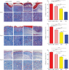
Collagen accumulation in PI wounds on days 5, 11, and 21. Masson staining shows that collagen deposition in the dermis was increased in ASC plus PRP-treated mice compared with the ASC group, PRP group, and controls. Collagen synthesis in wound skin. ×100 and ×200. P < 0.05, ∗∗P < 0.01, and ∗∗∗P < 0.001.
Figure 6
Figure 6
Effects of ASCs or PRP on wound neovascularization on days 5, 11, and 21 after cell or PRP transplantation. Immunohistochemical staining to reveal CD31 expression on days 5, 11, and 21 found more new blood vessels in tissue from ASC plus PRP-treated mice than from mice in the other groups. In all groups, fewer new blood vessels were seen on day 21 than on days 5 and 11. Formation of new vessels in PRP-treated and control mice on day 21 was not significantly different. ×200. P < 0.05, ∗∗P < 0.01, and ∗∗∗P < 0.001.
Figure 7
Figure 7
Effects of ASCs or PRP on wound neuroregeneration on days 11 and 21. Immunohistochemical staining of S100 in nerve cells in the dermis was increased more by ASCs plus PRP than by the other treatments. ×200. ∗∗P < 0.01, ∗∗∗P < 0.001.
Figure 8
Figure 8
ASCs tracked in vivo in mice with PI wounds. Accumulation of GFP-labelled ASCs at the wound margin on day 21 after transplantation of ASCs and ASCs combined with PRP. ×100.

Similar articles

Cited by

References

    1. Black J., Baharestani M., Cuddigan J., et al. National Pressure Ulcer Advisory Panel’s updated pressure ulcer staging system. Dermatology Nursing. 2007;19(4):343–349. quiz 50. - PubMed
    1. White-Chu E. F., Flock P., Struck B., Aronson L. Pressure ulcers in long-term care. Clinics in Geriatric Medicine. 2011;27(2):241–258. doi: 10.1016/j.cger.2011.02.001. - DOI - PubMed
    1. Cao Y., Krause J. S., DiPiro N. Risk factors for mortality after spinal cord injury in the USA. Spinal Cord. 2013;51(5):413–418. doi: 10.1038/sc.2013.2. - DOI - PubMed
    1. Krause J. S., Carter R. E. Risk of mortality after spinal cord injury: relationship with social support, education, and income. Spinal Cord. 2009;47(8):592–596. doi: 10.1038/sc.2009.15. - DOI - PMC - PubMed
    1. Demarre L., Van Lancker A., Van Hecke A., et al. The cost of prevention and treatment of pressure ulcers: a systematic review. International Journal of Nursing Studies. 2015;52(11):1754–1774. doi: 10.1016/j.ijnurstu.2015.06.006. - DOI - PubMed