

Torsion of a Large Myomatous Uterus Associated with Progressive Renal Failure and Paralytic Ileus in an 86-Year-Old Woman
- PMID: 31781441
- PMCID: PMC6875423
- DOI: 10.1155/2019/1601368
Torsion of a Large Myomatous Uterus Associated with Progressive Renal Failure and Paralytic Ileus in an 86-Year-Old Woman
Abstract
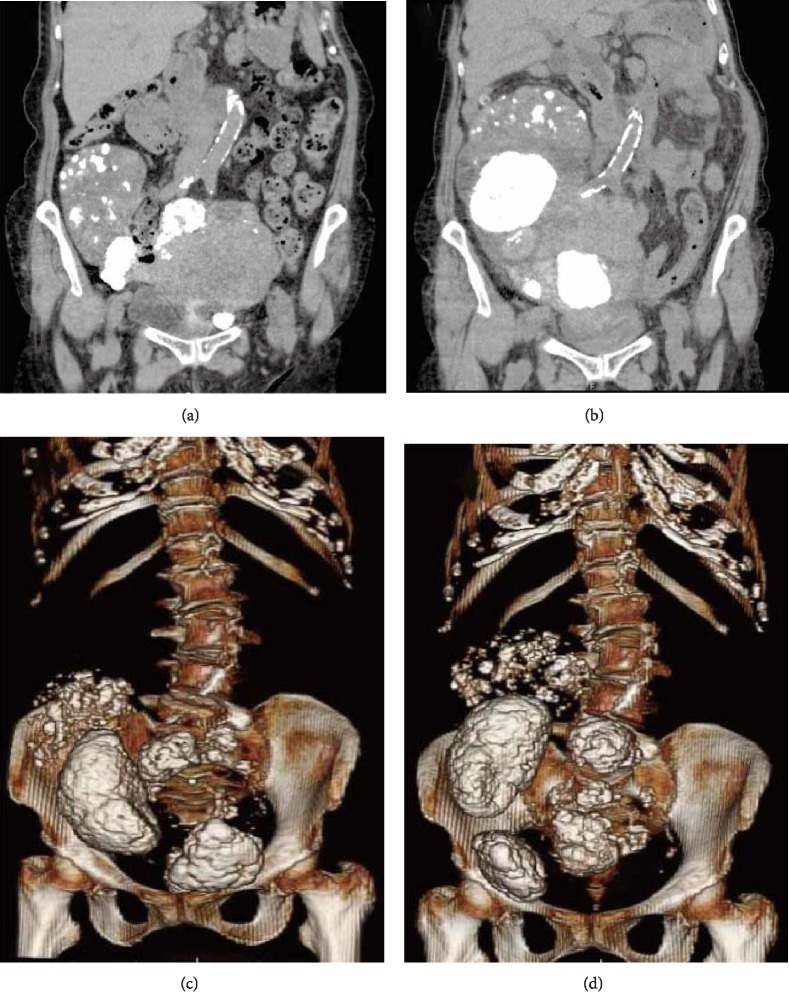
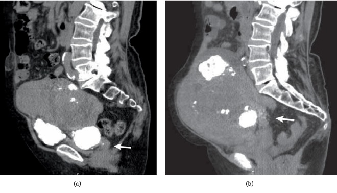
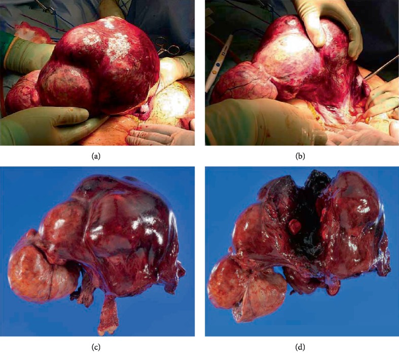
Uterine torsion of a nongravid uterus is rare, and proper diagnosis is challenging. Herein, we report a case of torsion of a large myomatous uterus in an 86-year-old woman who was presented with progressive renal failure and paralytic ileus. She was presented with abdominal discomfort, loss of appetite, and oliguria. A large myomatous uterus with broad calcification was identified when she underwent surgery to repair an umbilical hernia one year before the symptoms developed. Computed tomography revealed that one year later, the myomatous uterus significantly increased in size and the calcified lesion of the fibroid was largely displaced. She was also presented with paralytic ileus, and her general condition progressively worsened. Her serum creatinine levels were increased (3.5 mg/dL) and hemoglobin levels were low (8.5 g/dL). Emergency laparotomy revealed that the uterus was rotated 360 degrees clockwise at the level of the isthmus. The uterus was discolored, appearing dark red, and accompanied by broad congestion, and the cervix was elongated. The patient's renal function and ileus recovered after a hysterectomy. In conclusion, torsion of a large myomatous uterus could become life-threatening in an oldest-old woman, and early release of the torsion is necessary to avoid serious complications.
Copyright © 2019 Guiwen Wang et al.
Conflict of interest statement
The authors declare that they have no conflicts of interest regarding the publication of this paper.
Figures
Similar articles
-
Uterine Torsion in an Elderly Woman Associated with Leiomyoma and Continuously Elevating Muscle Enzymes: A Case Study and Review of Literature.Case Rep Obstet Gynecol. 2020 Oct 20;2020:8857300. doi: 10.1155/2020/8857300. eCollection 2020. Case Rep Obstet Gynecol. 2020. PMID: 33133709 Free PMC article.
-
Total Laparoscopic Hysterectomy and Bilateral Salpingo-Oophorectomy for a 6095-g Myomatous Uterus in a Patient of the Jehovah's Witness Faith.J Minim Invasive Gynecol. 2019 Jan;26(1):25-28. doi: 10.1016/j.jmig.2018.02.018. Epub 2018 Mar 5. J Minim Invasive Gynecol. 2019. PMID: 29518583
-
Torsion of a nongravid uterus with a large ovarian cyst: usefulness of contrast MR image.Gynecol Obstet Invest. 2007;63(3):163-5. doi: 10.1159/000096901. Epub 2006 Nov 9. Gynecol Obstet Invest. 2007. PMID: 17106201
-
Uteroenteric fistula resulting from fibroid expulsion after uterine fibroid embolization: case report and review of the literature.Cardiovasc Intervent Radiol. 2012 Oct;35(5):1231-6. doi: 10.1007/s00270-011-0318-4. Epub 2011 Dec 10. Cardiovasc Intervent Radiol. 2012. PMID: 22159908 Review.
-
Torsion of a non-gravid leiomyomatous uterus in a patient with myotonic dystrophy complaining of acute urinary retention: anaesthetic management for total abdominal hysterectomy.Clin Exp Obstet Gynecol. 2003;30(2-3):147-50. Clin Exp Obstet Gynecol. 2003. PMID: 12854863 Review.
Cited by
-
A Case Report of Uterine Torsion in a Postmenopausal Female with a Large Leiomyoma.J Radiol Case Rep. 2024 Jun 1;18(1):1-7. doi: 10.3941/jrcr.v18i1.5035. eCollection 2024. J Radiol Case Rep. 2024. PMID: 38910588 Free PMC article.
-
Uterine torsion in non-gravid women: A case report and review of cases reported in the last 20 years.SAGE Open Med Case Rep. 2021 Dec 22;9:2050313X211066649. doi: 10.1177/2050313X211066649. eCollection 2021. SAGE Open Med Case Rep. 2021. PMID: 34987820 Free PMC article.
-
Uterine Torsion in an Elderly Woman Associated with Leiomyoma and Continuously Elevating Muscle Enzymes: A Case Study and Review of Literature.Case Rep Obstet Gynecol. 2020 Oct 20;2020:8857300. doi: 10.1155/2020/8857300. eCollection 2020. Case Rep Obstet Gynecol. 2020. PMID: 33133709 Free PMC article.
-
Asymptomatic Cervical Amputation Caused by Uterine Torsion in a Non-Gravid Woman.J Clin Med. 2024 Dec 3;13(23):7356. doi: 10.3390/jcm13237356. J Clin Med. 2024. PMID: 39685814 Free PMC article.
References
-
- Common Toxicity Criteria. Version2.0. 1999. http://ctep.cancer.gov/protocolDevelopment/electronic_applications/docs/....
Publication types
LinkOut - more resources
Full Text Sources
