

Giant Squamous Cell Papilloma of the Eyelid-Diagnostic and Therapeutic Challenges
- PMID: 31781447
- PMCID: PMC6875255
- DOI: 10.1155/2019/5830493
Giant Squamous Cell Papilloma of the Eyelid-Diagnostic and Therapeutic Challenges
Abstract
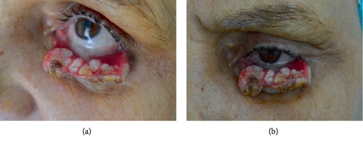
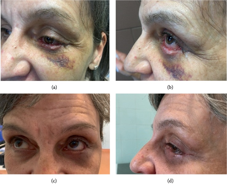
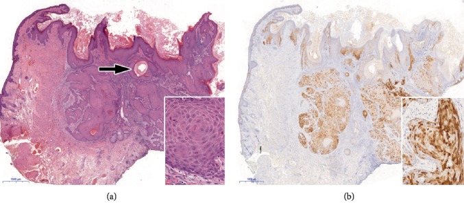
Squamous cell papilloma (SCP) is generally a human papillomavirus (HPV) induced exophytic or endophytic proliferation on the surface of the skin, oral cavity, larynx, esophagus, cervix, vagina, and anal canal. The endophytic type SCP can cause differential diagnostic difficulties with keratoacanthoma, inverted follicular keratosis, and squamous cell carcinoma; however, these lesions are not associated with HPV infection. The authors present a female patient who noticed an extremely rapidly growing tumor destructing the left lower eyelid. The histological analysis of the biopsy sample revealed a virus-induced squamoproliferative lesion. The eyelid affected was completely removed, and the histological examination resulted in a HPV induced endophytic squamous cell papilloma. The tarsus and the conjunctiva were replaced by a chondromucosal graft harvested from the nasal septum, while the skin defect could be closed directly. Restoration of the eyelid function has been achieved with satisfying functional and cosmetic results.
Copyright © 2019 Attila Vass et al.
Conflict of interest statement
The authors declare that they have no conflicts of interest.
Figures
Similar articles
-
Eyelid Papilloma.2023 Jul 4. In: StatPearls [Internet]. Treasure Island (FL): StatPearls Publishing; 2025 Jan–. 2023 Jul 4. In: StatPearls [Internet]. Treasure Island (FL): StatPearls Publishing; 2025 Jan–. PMID: 29262072 Free Books & Documents.
-
Conjuctival lesions - the relationship of papillomas and squamous cell carcinoma to HPV infection.Cesk Slov Oftalmol. 2018 Winter;74(3):92-97. doi: 10.31348/2018/1/2-3-2018. Cesk Slov Oftalmol. 2018. PMID: 30650971 English.
-
HPV type 16 in conjunctival and junctional papilloma, dysplasia, and squamous cell carcinoma.J Clin Pathol. 1995 Dec;48(12):1106-10. doi: 10.1136/jcp.48.12.1106. J Clin Pathol. 1995. PMID: 8567996 Free PMC article.
-
Basaloid squamous cell carcinoma arising in an inverted papilloma in the nasal cavity: A case report and review.Auris Nasus Larynx. 2017 Oct;44(5):624-628. doi: 10.1016/j.anl.2016.09.005. Epub 2016 Oct 7. Auris Nasus Larynx. 2017. PMID: 27720480 Review.
-
Human papillomavirus in sinonasal papillomas and squamous cell carcinoma.Laryngoscope. 1992 Sep;102(9):973-6. doi: 10.1288/00005537-199209000-00003. Laryngoscope. 1992. PMID: 1325585 Review.
Cited by
-
Nail Squamous Cell Papilloma: A Rare Case Report.Skin Appendage Disord. 2024 Apr;10(2):140-143. doi: 10.1159/000535080. Epub 2024 Jan 17. Skin Appendage Disord. 2024. PMID: 38572188 Free PMC article.
References
-
- Grayson W. Infectious diseases of the skin. In: Calonje J. E., Brenn T., Lazar A., McKee P., editors. McKee’s Pathology of the Skin. 4th. Vol. 1. Edinburgh: Elsevier/Saunders; 2011. pp. 760–895.
Publication types
LinkOut - more resources
Full Text Sources
