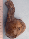
Somatic Malignant Transformation of a Testicular Teratoma: A Case Report and an Unusual Presentation
- PMID: 31781458
- PMCID: PMC6874974
- DOI: 10.1155/2019/5273607
Somatic Malignant Transformation of a Testicular Teratoma: A Case Report and an Unusual Presentation
Abstract
Testicular cancer represents 1% of all malignant tumors in men. About 95% of testicular cancers are germ cell tumors (GCTs). These can be divided into nonseminomatous GCTs (NSGCTs) and seminomas. NSGCTs include teratomas, yolk sac tumors, embryonal carcinomas, choriocarcinomas, and mixed tumors. Only 2-6% of testicular teratomas are pure teratomas. Pure teratomas can be subdivided into prepubertal and postpubertal. The prognosis is significantly different between these two age groups. Different from teratomas in ovary, the immaturity in a teratoma is not an indication of their biologic behavior; the age of the patient is of greater importance. Malignant transformation of teratoma occurs in only 3-6% of testicular GCTs. The most frequent transformed histologic types consist of rhabdomyosarcoma, adenocarcinoma, and primitive neuroectodermal tumors. We report a rare case of pure postpubertal testicular teratoma with a secondary somatic malignancy that was an incidental finding in a patient presenting with lower back pain and testicular torsion.
Copyright © 2019 Dalia Y. Ibrahim and Hongliu Sun.
Conflict of interest statement
The authors declare that they have no conflicts of interest.
Figures


Similar articles
-
Germ cell tumors of the gonads: a selective review emphasizing problems in differential diagnosis, newly appreciated, and controversial issues.Mod Pathol. 2005 Feb;18 Suppl 2:S61-79. doi: 10.1038/modpathol.3800310. Mod Pathol. 2005. PMID: 15761467 Review.
-
[Studies on nuclear DNA content in testicular germ cell tumors using flow cytometry].Hokkaido Igaku Zasshi. 1992 Nov;67(6):801-14. Hokkaido Igaku Zasshi. 1992. PMID: 1336479 Japanese.
-
A Clinicopathologic and Molecular Analysis of 34 Mediastinal Germ Cell Tumors Suggesting Different Modes of Teratoma Development.Am J Surg Pathol. 2018 Dec;42(12):1662-1673. doi: 10.1097/PAS.0000000000001164. Am J Surg Pathol. 2018. PMID: 30256256
-
Pathology and molecular biology of teratomas in childhood and adolescence.Klin Padiatr. 2006 Nov-Dec;218(6):296-302. doi: 10.1055/s-2006-942271. Klin Padiatr. 2006. PMID: 17080330
-
Secondary malignant transformation of testicular teratomas: case series and literature review.Actas Urol Esp. 2014 Nov;38(9):622-7. doi: 10.1016/j.acuro.2014.02.014. Epub 2014 Jun 6. Actas Urol Esp. 2014. PMID: 24909334 Review. English, Spanish.
Cited by
-
Testicular non-seminomatous mixed germ cell tumor with rhabdomyosarcoma and retroperitoneal metastatic mass: A case report.Urol Case Rep. 2020 Oct 28;34:101475. doi: 10.1016/j.eucr.2020.101475. eCollection 2021 Jan. Urol Case Rep. 2020. PMID: 33294376 Free PMC article.
-
Metastatic Primary Testicular Neuroendocrine Carcinoma Associated with Somatic Malignant Transformation of Teratoma: A Rare Case Report.Case Rep Oncol. 2022 Mar 10;15(1):191-198. doi: 10.1159/000521998. eCollection 2022 Jan-Apr. Case Rep Oncol. 2022. PMID: 35431867 Free PMC article.
-
Unveiling the genomic landscape of possible metastatic malignant transformation of teratoma secondary to cisplatin-chemotherapy: a Tempus gene analysis-based case report literature review.Front Oncol. 2023 Jun 22;13:1192843. doi: 10.3389/fonc.2023.1192843. eCollection 2023. Front Oncol. 2023. PMID: 37427132 Free PMC article.
-
Calcified metastases of teratoma.J Bras Pneumol. 2021 Jan 20;47(1):e20200462. doi: 10.36416/1806-3756/e20200462. J Bras Pneumol. 2021. PMID: 33503135 Free PMC article. No abstract available.
-
Low level expression of human telomerase reverse transcriptase predicts cancer-related death and progression in embryonal carcinoma.J Cancer Res Clin Oncol. 2020 Nov;146(11):2753-2775. doi: 10.1007/s00432-020-03319-2. Epub 2020 Jul 17. J Cancer Res Clin Oncol. 2020. PMID: 32681293 Free PMC article.
References
-
- Şekerci Ç. A., Yılmaz G., Yılmaz I. B. Pure testicular cystic teratoma in an adult patient. Journal of Urological Surgery. 2015;2(4):214–215. doi: 10.4274/jus.207. - DOI
-
- Kudva R., Kudva A. Pure testicular teratoma presenting as a metastatic germ cell tumor. International Journal of Scientific and Research Publications. 2014;4(3):1–4.
-
- Williamson S. R., Delahunt B., Magi-Galluzzi C., et al. The World Health Organization 2016 classification of testicular germ cell tumours: a review and update from the international society of urological pathology testis consultation panel. Histopathology. 2017;70(3):335–346. doi: 10.1111/his.13102. - DOI - PubMed
Publication types
LinkOut - more resources
Full Text Sources

