Case Reports
. 2019 Nov 12:2019:5416936.
doi: 10.1155/2019/5416936. eCollection 2019.

Renal Tubular Dysgenesis in a Case of Fetus Acardius Amorphus

Affiliations
Case Reports

Renal Tubular Dysgenesis in a Case of Fetus Acardius Amorphus

C Thoeni et al. Case Rep Pathol. .

Abstract

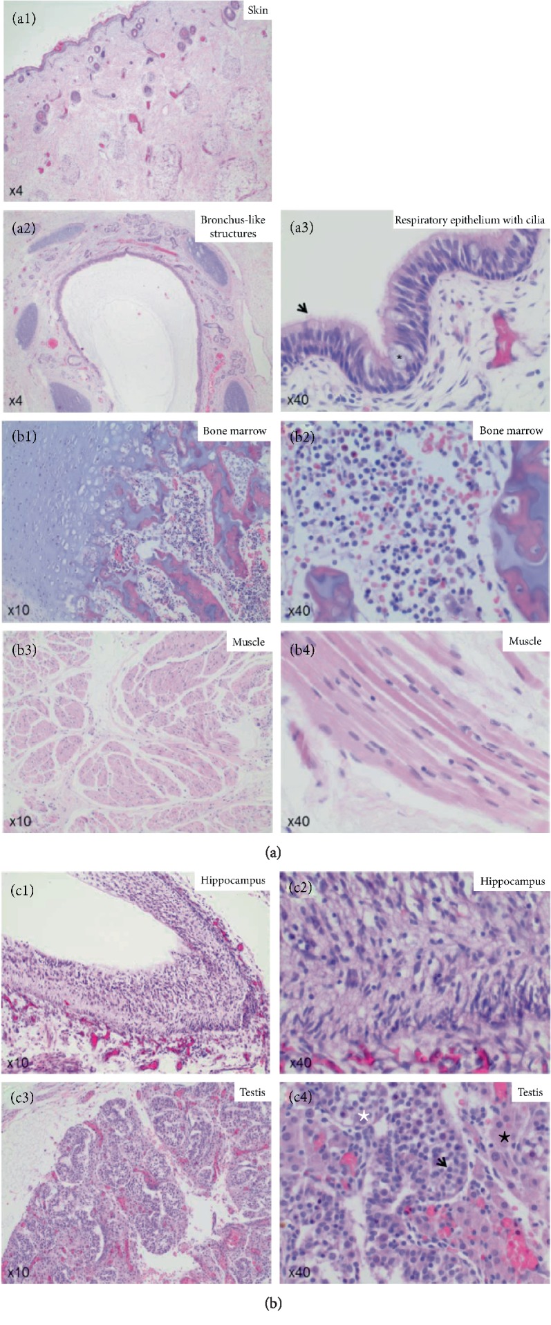
Fetus acardius amorphus is a rare congenital malformation characterized by the lack of a functional heart, the presence of a bivascular umbilical cord, as well as a developed and organized skeletal system and partially organized inner organs. Fetus acardii mostly occur in multiple gestations. The pathogenesis of this entity is not clarified yet. It has been hypothesized that, although formation of anastomosing vessels between the co-twin and the anomalous embryo as well as reverse directed blood flow within the umbilical arteries of the weaker twin may allow sufficient blood flow to form rudimentary internal organs, it is insufficient to develop a fully functional heart. We had a case of fetus acardius amorphus, where we performed autopsy as well as routine histology assessment to identify different types of tissues. We showed that our fetus acardius amorphus demonstrated histomorphological features of renal tubular dysgenesis, confirmed by lack of proximal tubules, extramedullary hematopoiesis and increased number of smooth muscle actin positive vessels. This is a novel finding and has not been reported previously.


Conflict of interest statement

The authors declare that they have no conflicts of interest.

Figures

Figure 1
Figure 1
(a) Photographs of the Fetus acardius amorphus. Fetus acardius amorphus shown as a rounded ovoid mass with a small pedicle symbolizing the umbilical cord. On one surface there exists a colloidal mass, as well as a short pedicle with little hair on it. (b) The Center of Fetus acardius amorphus illustrates an organized bone structure predicted to be the vertebra column, surrounded by yellowish tissue as well as red masses. (c1 and c2) Facitron X-ray analysis of the skeletal system of the Fetus acardius amorphus. The Fetus acardius amorphus shows a complete organized vertebral column, with a rudimentary rib cage and an ovoid bone structure (white arrow) without axial organization of a head, limbs or cranial-caudal poles. (d1 and d2) CISH analysis of interphase cells within paraffin sections of male gonadal tissue using the CEN X/Y Probe. CEN X/Y Probe hybridizes on regular male interphase cells demonstrated by one red (chromosome X) and one green (chromosome Y) signal per nucleus. The Fetus acardius amorphus shows a male XY genotype. Scale bar = 20 μm (D1), scale bar = 5 μm (D2).
Figure 2
Figure 2
(a1): Hematoxylin and eosin (HE) staining of the skin of the Fetus acardius amorphus. Fetus acardius amorphus shows differentiated skin tissue, with a stratum corneum, an epidermis, a dermis and a subcutis. Images are shown in 4x magnification. (a2 and a3) Hematoxylin and Eosin (HE) staining of the core mass of the Fetus acardius amorphus. Fetus acardius amorphus shows organized bronchus-like structures with respiratory epithelium, goblet cells and cilia (black arrow) on the surface. Images are shown in 4x magnification (a2) and in 40x magnification (a3). (b1 and b2) Hematoxylin and Eosin (HE) staining of the core mass of the Fetus acardius amorphus. Fetus acardius amorphus shows differentiated bone and cartilage with bone marrow cells like megakaryocytes, erythrocyte precursor cells as well as myelocytes in between. Images are shown in 10x magnification (b1) and in 40x magnification (b2). (b3 and b4) Hematoxylin and Eosin (HE) staining of the core mass of the Fetus acardius amorphus. Fetus acardius amorphus shows differentiated skeletal muscle with long, shapoid myocytes with an ovoid nucleus in the center. Images are shown in 10x magnification (b3) and in 40x magnification (b4). (c1 and c2) Hematoxylin and Eosin (HE) staining of the cyst-like structure in the Fetus acardius amorphus. Fetus acardius amorphus shows differentiated neurons of the hippocampus. Images are shown in 10x magnification (c1) and in 40x magnification (c2). (c3 and c4) Hematoxylin and Eosin (HE) staining of the gonadal glands in the Fetus acardius amorphus. Fetus acardius amorphus shows gonadal glands with well-formed seminiferous tubules showing Ledyig cells between (black star), and spermatogonia (black arrow) and Sertoli cells (white star) inside. Images are shown in 10x magnification (c3) and in 40x magnification (c4).
Figure 3
Figure 3
Hematoxylin and Eosin (HE) staining and immunohistochemistry staining for villin, mucin 2 and calretinin in the intestine of the Fetus acardius amorphus. Villin is stained in red (ALEXA 568), mucin 2 in green (ALEXA 488) and nuclei in blue (Hoechst). Fetus acardius shows differentiated small (a) and large (b) intestine with positive apical villin staining (red) marking the intestinal brush border in cells of the crypt and villi and mucin 2 (green) positive Goblet cells in crypts and villi. Ganglion cells of the Auerbach and Meissner Plexus were also present shown by positive Calretinin staining. Images are shown in 10x and in 20x magnifications.
Figure 4
Figure 4
(a) PAS staining and immunohistochemistry staining for villin of fetal renal tissue. In immunohistochemistry, villin is stained in red (ALEXA 568) and nuclei in blue (Hoechst). Fetus acardius shows differentiated kidney with cortex and medulla, but lack of proximal tubules as shown by negative villin staining. Additionally, distal tubules of the fetus acardius amorphus show mild nephrocalcinosis (white star). PT (proximal tubule), G (glomerulus). Images are shown in 10x, 20x and 40x magnifications. (b) Hematoxylin and Eosin (HE) staining and immunohistochemistry staining for MAC 387 staining in fetal renal tissue. MAC 387 positive cells were detected in the interstitial tissue of the kidney in the Fetus acardius amorphus, those cells are marked with a black arrow in the Hematoxylin and Eosin (HE) staining labeling erythrocyte precursor cells. MAC 387 is stained in red (ALEXA 568), nuclei stained with in blue (Hoechst). Images are shown in 20x magnification. (c) CD31 immunohistochemistry staining in fetal renal tissue. CD31 (brown) is staining vessels in the renal cortex and medulla, nuclei are stained with hematoxylin in blue. Increased number of CD31 positive vessels is detected, in particular in the renal cortex of the fetus acardius amorphus.Images are shown in 10x magnification. (d) Smooth muscle actin (SMA) staining in fetal renal tissue. Increased number of SMA positive vessels in particular the cortex of the fetus acardius amorphus. SMA is stained in green, nuclei stained with Hoechst in blue. Glomeruli are marked with a white star, tubules with white arrows. Images are shown in 10x magnification.

Similar articles

Cited by

  • A complementary study approach unravels novel players in the pathoetiology of Hirschsprung disease.
    Mederer T, Schmitteckert S, Volz J, Martínez C, Röth R, Thumberger T, Eckstein V, Scheuerer J, Thöni C, Lasitschka F, Carstensen L, Günther P, Holland-Cunz S, Hofstra R, Brosens E, Rosenfeld JA, Schaaf CP, Schriemer D, Ceccherini I, Rusmini M, Tilghman J, Luzón-Toro B, Torroglosa A, Borrego S, Sze-Man Tang C, Garcia-Barceló M, Tam P, Paramasivam N, Bewerunge-Hudler M, De La Torre C, Gretz N, Rappold GA, Romero P, Niesler B. Mederer T, et al. PLoS Genet. 2020 Nov 5;16(11):e1009106. doi: 10.1371/journal.pgen.1009106. eCollection 2020 Nov. PLoS Genet. 2020. PMID: 33151932 Free PMC article.

References

    1. Harvey. Acephalous monster at the full period of pregnancy. Lancet. 1848;ii:696
    1. Spliedt. Anatomical description of an acardiac monstrosity. British and foreign medico-chirurgical review. 1860;26:543.
    1. Slyman W. An acephalous acardiac monster of six months’ gestation with rudimentary heart. Transactions of the Obstetric Society of London. 1889;31:258–262.
    1. Stewart M. J. A specimen of foetus acardiacus amorphus. Proceedings of the Royal Society Medicine 7 (Obstet Gynaecol Sect) 1914:131–138. - PMC - PubMed
    1. Wolff B. Über eine Drillingsgeburt mit einem acardius. Archiv für Gynäkologie. 1971;59(2):294–313. doi: 10.1007/BF01833524. - DOI

Publication types

LinkOut - more resources