

Case Reports
doi: 10.1155/2019/8049393.
eCollection 2019.
Abdominal Abscesses and Destruction of Inguinal Canal with Mesh Dislocation caused by a Perforated Diverticulitis
Affiliations
- PMID: 31781467
- PMCID: PMC6874982
- DOI: 10.1155/2019/8049393
Item in Clipboard
Case Reports
Abdominal Abscesses and Destruction of Inguinal Canal with Mesh Dislocation caused by a Perforated Diverticulitis
Case Rep Surg.
.
Abstract
The diverticulitis is a frequent disease of the gastrointestinal tract. It may lead to a variety of severe complications. In some cases, it has to be surgically treated. Herein, we present a rare case of a 66-year-old man, who suffered from a painful, visible "fist sized" mass of the left lower abdomen. A perforated diverticulitis with abdominal, cutaneous abscesses and destruction of the inguinal canal with mesh dislocation was diagnosed and successfully surgically treated.
Copyright © 2019 Christoph Paasch et al.
Conflict of interest statement
The authors declare that they have no conflicts of interest.
Figures

The picture shows a “fist sized” mass of the left lower abdomen.

Computed tomography of the abdomen; the picture highlights an inflamed 4.6 × 9.5 cm sized conglomerate adjacent to the abdominal wall and the left inguinal canal (yellow and white dashed arrow). The prior implanted mesh seemed to be partially dislocated to the abdominal cavity (white star).

The picture shows the opened left inguinal canal with the mesh (white point). On the right side of the image, the mesh has been removed.
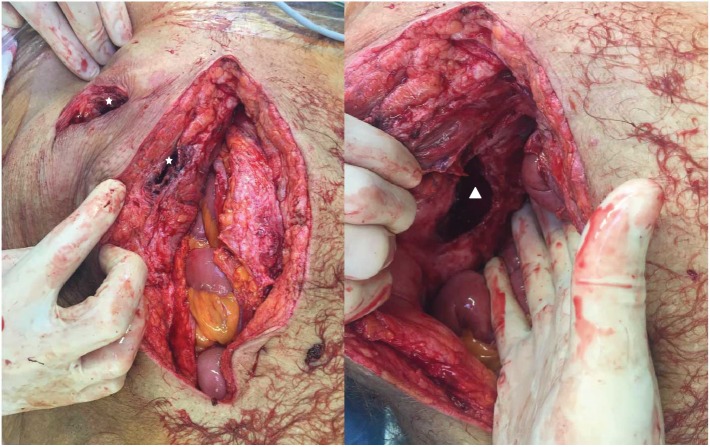
The subcutaneous and cutaneous opened abscess cavity is indicated by the white star. This cavity was connected with the left inguinal canal (white triangle).
Similar articles
-
Subcutaneous emphysema: a rare manifestation of a perforated diverticulitis in a patent inguinal canal.Hernia. 2007 Jun;11(3):261-3. doi: 10.1007/s10029-006-0172-5. Epub 2006 Nov 29. Hernia. 2007. PMID: 17136307
-
[Multiple recurrent perforated jejunal diverticulitis].Chirurg. 2002 Dec;73(12):1218-20. doi: 10.1007/s00104-002-0538-x. Chirurg. 2002. PMID: 12491052 German.
-
Abdominal wall abscess associated with perforated jejunal diverticulitis: report of a case.Surg Today. 2005;35(8):682-6. doi: 10.1007/s00595-004-2972-5. Surg Today. 2005. PMID: 16034551
-
Ultrasound of colon diverticulitis.Dig Dis. 2012;30(1):56-9. doi: 10.1159/000336620. Epub 2012 May 3. Dig Dis. 2012. PMID: 22572686 Review.
-
Operative Strategies for Perforated Diverticulitis: A Systematic Review and Meta-analysis.Dis Colon Rectum. 2018 Dec;61(12):1442-1453. doi: 10.1097/DCR.0000000000001149. Dis Colon Rectum. 2018. PMID: 30371549
Cited by
-
Early sigmoid perforation involving left inguinal hernia mesh repair: a case report.Ann Med Surg (Lond). 2023 Feb 7;85(2):181-183. doi: 10.1097/MS9.0000000000000081. eCollection 2023 Feb. Ann Med Surg (Lond). 2023. PMID: 36845822 Free PMC article.
References
Publication types
LinkOut - more resources
Full Text Sources
