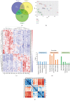
In-Depth Characterization of Mass Spectrometry-Based Proteomic Profiles Revealed Novel Signature Proteins Associated with Liver Metastatic Colorectal Cancers
- PMID: 31781478
- PMCID: PMC6875276
- DOI: 10.1155/2019/7653230
In-Depth Characterization of Mass Spectrometry-Based Proteomic Profiles Revealed Novel Signature Proteins Associated with Liver Metastatic Colorectal Cancers
Abstract
Liver metastasis is the most common form of metastatic colorectal cancers during the course of the disease. The global change in protein abundance in liver metastatic colorectal cancers and its role in metastasis establishment have not been comprehensively analyzed. In the present study, fresh-frozen tissue samples including normal colon/localized/liver metastatic CRCs from each recruited patient were analyzed by quantitative proteomics using a multiplexed TMT labeling strategy. Around 5000 protein groups were quantified from all samples. The proteomic profile of localized/metastatic CRCs varied greatly from that of normal colon tissues; differential proteins were mainly from extracellular regions and participate in immune activities, which is crucial for the chronic inflammation signaling pathways in the tumor microenvironment. Further statistical analysis revealed 47 proteins exhibiting statistical significance between localized and metastatic CRCs, of which FILI1P1 and PLG were identified for the first time in proteomic data, which were highly associated with liver metastasis in CRCs.
Copyright © 2019 Xin Ku et al.
Conflict of interest statement
The authors declare no potential conflicts of interest.
Figures


Similar articles
-
In-depth characterization of the secretome of colorectal cancer metastatic cells identifies key proteins in cell adhesion, migration, and invasion.Mol Cell Proteomics. 2013 Jun;12(6):1602-20. doi: 10.1074/mcp.M112.022848. Epub 2013 Feb 26. Mol Cell Proteomics. 2013. PMID: 23443137 Free PMC article.
-
Immuno-proteomic discovery of tumor tissue autoantigens identifies olfactomedin 4, CD11b, and integrin alpha-2 as markers of colorectal cancer with liver metastases.J Proteomics. 2017 Sep 25;168:53-65. doi: 10.1016/j.jprot.2017.06.021. Epub 2017 Jun 29. J Proteomics. 2017. PMID: 28669815
-
Proteomics analysis of the matrisome from MC38 experimental mouse liver metastases.Am J Physiol Gastrointest Liver Physiol. 2019 Nov 1;317(5):G625-G639. doi: 10.1152/ajpgi.00014.2019. Epub 2019 Sep 23. Am J Physiol Gastrointest Liver Physiol. 2019. PMID: 31545917 Free PMC article.
-
Genomics and proteomics approaches for biomarker discovery in sporadic colorectal cancer with metastasis.Cancer Genomics Proteomics. 2013 Jan-Feb;10(1):19-25. Cancer Genomics Proteomics. 2013. PMID: 23382583 Review.
-
The cancer secretome, current status and opportunities in the lung, breast and colorectal cancer context.Biochim Biophys Acta. 2013 Nov;1834(11):2242-58. doi: 10.1016/j.bbapap.2013.01.029. Epub 2013 Jan 31. Biochim Biophys Acta. 2013. PMID: 23376433 Review.
Cited by
-
The Role of Biomarkers in the Management of Colorectal Liver Metastases.Cancers (Basel). 2022 Sep 22;14(19):4602. doi: 10.3390/cancers14194602. Cancers (Basel). 2022. PMID: 36230522 Free PMC article. Review.
-
Advancements in Oncoproteomics Technologies: Treading toward Translation into Clinical Practice.Proteomes. 2023 Jan 10;11(1):2. doi: 10.3390/proteomes11010002. Proteomes. 2023. PMID: 36648960 Free PMC article. Review.
-
Personalized Therapy and Liquid Biopsy-A Focus on Colorectal Cancer.J Pers Med. 2021 Jul 1;11(7):630. doi: 10.3390/jpm11070630. J Pers Med. 2021. PMID: 34357097 Free PMC article. Review.
-
Multilevel Heterogeneity of Colorectal Cancer Liver Metastasis.Cancers (Basel). 2023 Dec 21;16(1):59. doi: 10.3390/cancers16010059. Cancers (Basel). 2023. PMID: 38201487 Free PMC article. Review.
-
Proteomic Profiling and Biomarker Discovery in Colorectal Liver Metastases.Int J Mol Sci. 2022 May 29;23(11):6091. doi: 10.3390/ijms23116091. Int J Mol Sci. 2022. PMID: 35682769 Free PMC article. Review.
References
MeSH terms
Substances
LinkOut - more resources
Full Text Sources
Medical
Miscellaneous

