. 2019 Nov 8:7:296.
doi: 10.3389/fbioe.2019.00296. eCollection 2019.

Non-invasive Production of Multi-Compartmental Biodegradable Polymer Microneedles for Controlled Intradermal Drug Release of Labile Molecules

Affiliations

Non-invasive Production of Multi-Compartmental Biodegradable Polymer Microneedles for Controlled Intradermal Drug Release of Labile Molecules

Mario Battisti et al. Front Bioeng Biotechnol. .

Abstract

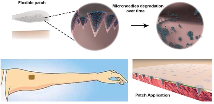
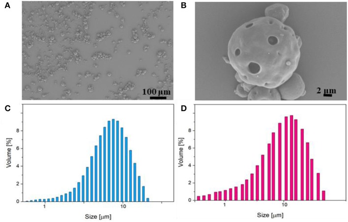
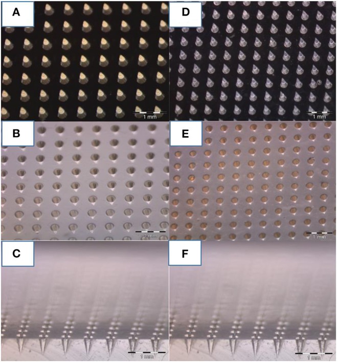
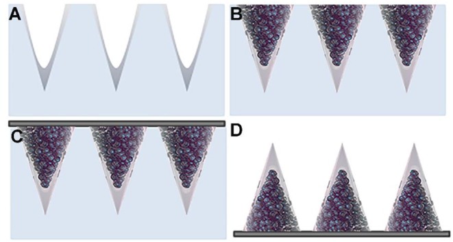
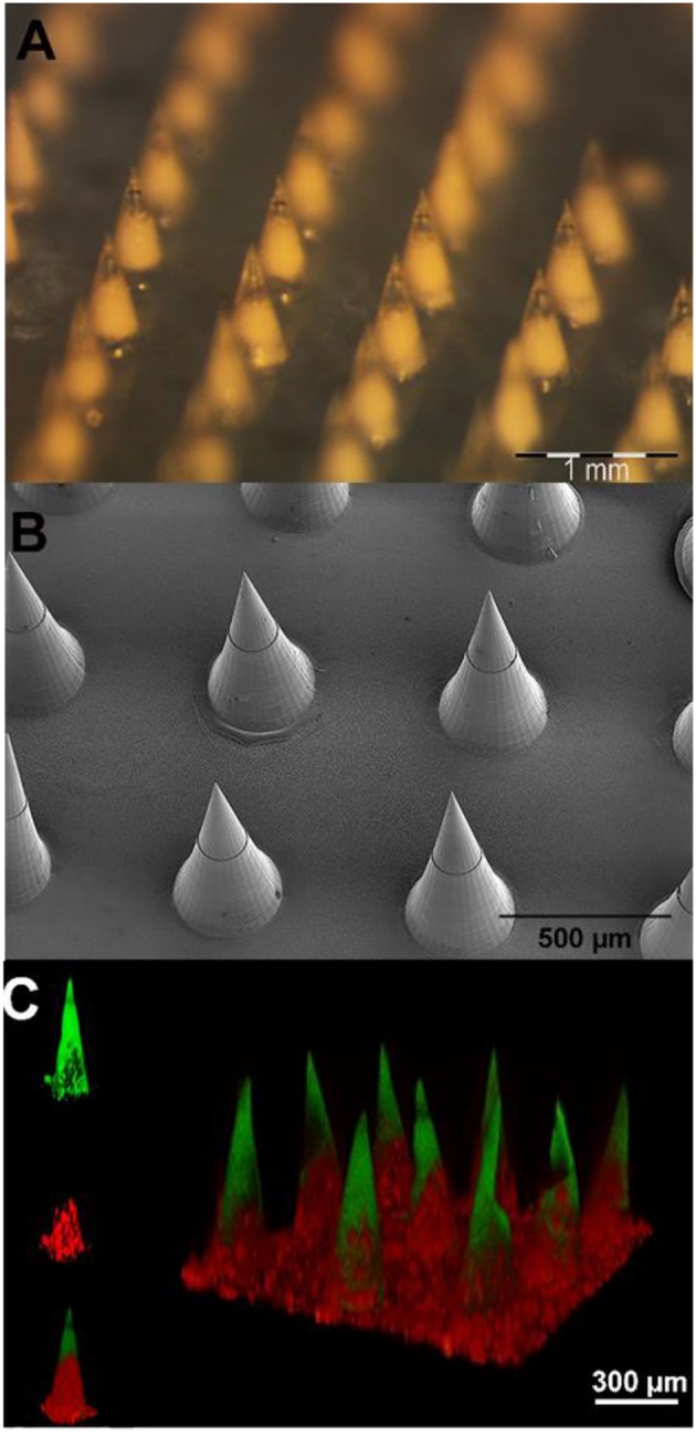
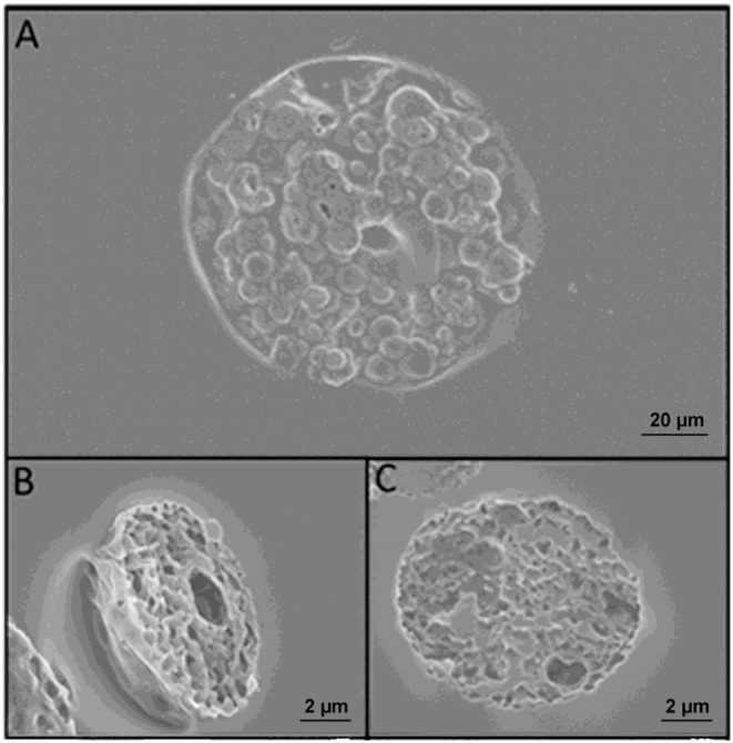
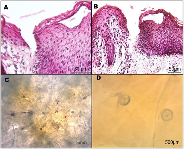
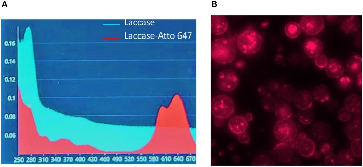
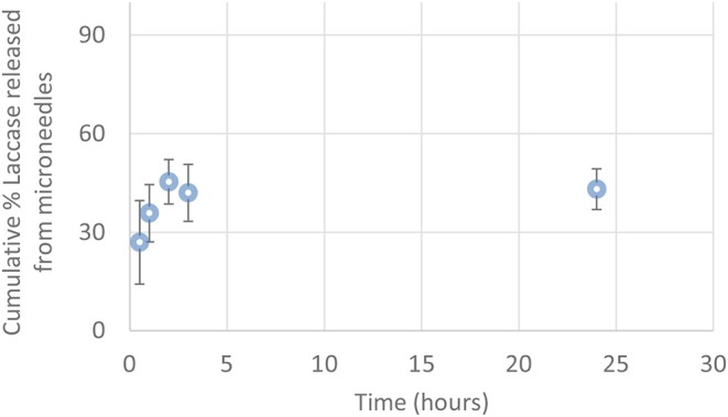
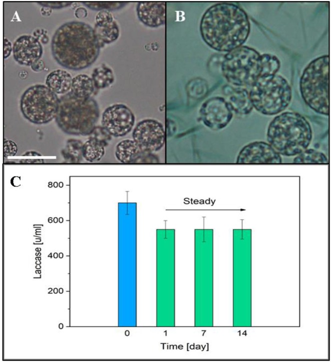
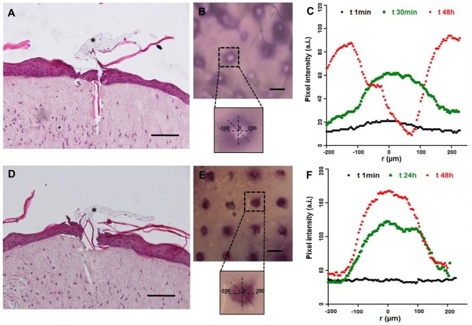
Transdermal drug delivery represents an appealing alternative to conventional drug administration systems. In fact, due to their high patient compliance, the development of dissolvable and biodegradable polymer microneedles has recently attracted great attention. Although stamp-based procedures guarantee high tip resolution and reproducibility, they have long processing times, low levels of system engineering, are a source of possible contaminants, and thermo-sensitive drugs cannot be used in conjunction with them. In this work, a novel stamp-based microneedle fabrication method is proposed. It provides a rapid room-temperature production of multi-compartmental biodegradable polymeric microneedles for controlled intradermal drug release. Solvent casting was carried out for only a few minutes and produced a short dissolvable tip made of polyvinylpyrrolidone (PVP). The rest of the stamp was then filled with degradable poly(lactide-co-glycolide) (PLGA) microparticles (μPs) quickly compacted with a vapor-assisted plasticization. The outcome was an array of microneedles with tunable release. The ability of the resulting microneedles to indent was assessed using pig cadaver skin. Controlled intradermal delivery was demonstrated by loading both the tip and the body of the microneedles with model therapeutics; POXA1b laccase from Pleurotus ostreatus is a commercial enzyme used for the whitening of skin spots. The action and indentation of the enzyme-loaded microneedle action were assessed in an in vitro skin model and this highlighted their ability to control the kinetic release of the encapsulated compound.

Keywords: controlled release; enzyme; multi-compartmental; polymer microneedles; skin model.


Figures

Graphical Abstract
Graphical Abstract
Highly controlled intradermal delivery can be performed with multi-compartmental polymer microneedles that are able to indent the skin and be implanted in it with a fast dissolving tip and a slow degradable body made of porous PLGA μPs.
Figure 1
Figure 1
(A,B) SEM images of enzyme-loaded μPs at different magnifications. Particle size distribution analysis of PLGA μPs performed by Mastersizer 3000. (C) Sulphorhodamine-loaded μPs. (D) Laccase-loaded μPs.
Figure 2
Figure 2
Optical image of microneedles molded at various steps. (A) Master of 600 um of height and 300 um of bases of microneedles fabricated by 2PP, (B) PDMS stamp replicated on the master, and (C) cross-section of the PDMS mold. (D) NOA master replicated on the PDMS stamp, and (E) final PDMS stamp replicated on it (F) including a cross-section. A comparison of photos (C,F) reveal how, despite the various stages of replication, the needles keep tips with the same curvature radius.
Figure 3
Figure 3
Schematic view of the multi-compartmental microneedle array. PDMS stamp with (A) PVP deposited by spin coating and with (B) PLGA μPs compacted and sintered. (C) Application of the harvesting layer on the stamp. (D) Microneedle array extracted from the stamp.
Figure 4
Figure 4
(A) Stereomicroscope micrographs of microparticle-loaded microneedle patches. (B) Scanning electron microscopy images of microneedle arrays. (C) 3D confocal reconstruction of a microneedle array. The FITC loaded tips are shown in green, while the sulphorhodamine B loaded PLGA μPs are in red.
Figure 5
Figure 5
(A) SEM images of microneedles slices obtained with cryosectioning. (B) Slice of a microparticle contained in the microneedle after sintering. (C) Slice of a microparticle just prepared. A comparison of (B,C) reveals that sintering does not change the morphology of the microparticles.
Figure 6
Figure 6
Microneedle patches efficiently penetrate the skin. (A,B) Cross-sectional images of the H/E stained skin after microneedle removal. (C,D) Stereomicroscope image of microneedles in the skin.
Figure 7
Figure 7
Laccase-Atto 647 μPs. (A) UV spectrum of Laccase (light blue line) and Laccase Atto647 (red line). (B) Fluorescence image of Laccase-Atto647 μPs.
Figure 8
Figure 8
Cumulative percentage of Laccase-Atto647 released from μPs in the microneedles (0.5–24 h) (n = 3).
Figure 9
Figure 9
(A) Optical image of μPs slice. (B) Optical μPs slice with a solution of ABTS and buffer. Scale bar 10 μm. (C) Laccase activity in μPs up to 14 days after the production. The blue column represents the initial enzyme activity used for μPs preparation.
Figure 10
Figure 10
Functional test of microneedles for transdermal delivery of high molecular weight substances in a full-thickness human skin model. The pictures in (A–C) refer to microneedle configuration 1, while (D–F) refer to configuration 2. (A,D) Histological images of Endo-HSE 48 h after microneedle indentation to highlight the transdermal penetration; black asterisks indicate the PVP polymer remaining after medical tape removal (scale bar = 100 μm). (B,E) Stereomicroscopic images of Endo-HSE 48 h after indentation (scale bar = 500 μm). The inserts of the stereomicroscopic images are the schematic representation of the methods used to calculate the diffusive radius. (C,F) The graphs plot the pixel intensity at three time points correlated to the concentration of the ABTS oxidation product diffusing into ECM vs. the radius of the diffusion pattern.

References

    1. Arya J. M., Dewitt K., Scott-Garrard M., Chiang Y.-W., Prausnitz M. R. (2016). Rabies vaccination in dogs using a dissolving microneedle patch. J. Control. Release 239, 19–26. 10.1016/j.jconrel.2016.08.012 - DOI - PubMed
    1. Bellas E., Seiberg M., Garlick J., Kaplan D. L. (2012). In vitro 3D full-thickness skin-equivalent tissue model using silk and collagen biomaterials. Macromol. Biosci. 12, 1627–1636. 10.1002/mabi.201200262 - DOI - PMC - PubMed
    1. Bobo D., Robinson K. J., Islam J., Thurecht K. J., Corrie S. R. (2016). Nanoparticle-based medicines: a review of FDA-approved materials and clinical trials to date. Pharm. Res. 33, 2373–2387. 10.1007/s11095-016-1958-5 - DOI - PubMed
    1. Bouwstra J. A., Ponec M. (2006). The skin barrier in healthy and diseased state. Biochim. Biophys. Acta 1758, 2080–2095. 10.1016/j.bbamem.2006.06.021 - DOI - PubMed
    1. Bronaugh R., Maibach H. (1999). Percutaneous absorption drug-cosmetics-mechanism-methodology. J. Appl. Cosmetol. 17, 116–119.

LinkOut - more resources