Exploring the Biomarkers of Sepsis-Associated Encephalopathy (SAE): Metabolomics Evidence from Gas Chromatography-Mass Spectrometry
- PMID: 31781604
- PMCID: PMC6875220
- DOI: 10.1155/2019/2612849
Exploring the Biomarkers of Sepsis-Associated Encephalopathy (SAE): Metabolomics Evidence from Gas Chromatography-Mass Spectrometry
Abstract
Background: Sepsis-associated encephalopathy (SAE) is a transient and reversible brain dysfunction, that occurs when the source of sepsis is located outside of the central nervous system; SAE affects nearly 30% of septic patients at admission and is a risk factor for mortality. In our study, we sought to determine whether metabolite changes in plasma could be a potential biomarker for the early diagnosis and/or the prediction of the prognosis of sepsis.
Method: A total of 31 SAE patients and 28 healthy controls matched by age, gender, and body mass index (BMI) participated in our study. SAE patients were divided into four groups according to the Glasgow Coma Score (GCS). Plasma samples were collected and used to detect metabolism changes by gas chromatography-mass spectrometry (GC-MS). Analysis of variance was used to determine which metabolites significantly differed between the control and SAE groups.
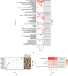
Results: We identified a total of 63 metabolites that showed significant differences among the SAE and control groups. In particular, the 4 common metabolites in the four groups were 4-hydroxyphenylacetic acid; carbostyril, 3-ethyl-4,7-dimethoxy (35.8%); malic acid peak 1; and oxalic acid. The concentration of 4-hydroxyphenylacetic acid in sepsis patients decreased with a decrease of the GCS.
Conclusions: According to recent research on SAE, metabolic disturbances in tissue and cells may be the main pathophysiology of this condition. In our study, we found a correlation between the concentration of 4-hydroxyphenylacetic acid and the severity of consciousness disorders. We suggest that 4-hydroxyphenylacetic acid may be a potential biomarker for SAE and useful in predicting patient prognosis.
Copyright © 2019 Jing Zhu et al.
Conflict of interest statement
The authors declare that they have no conflicts of interest.
Figures


References
MeSH terms
Substances
LinkOut - more resources
Full Text Sources
Medical
Miscellaneous

