Review
. 2019 Nov 12:2019:7453260.
doi: 10.1155/2019/7453260. eCollection 2019.

Immunological Aspects of Graves' Ophthalmopathy

Affiliations
Review

Immunological Aspects of Graves' Ophthalmopathy

Dominika Łacheta et al. Biomed Res Int. .

Abstract

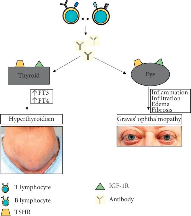
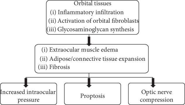
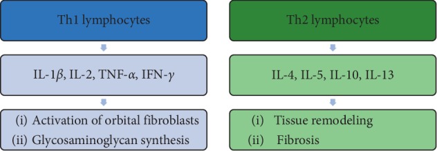
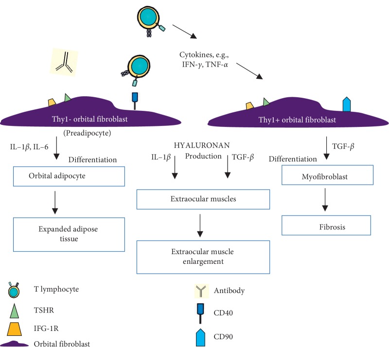
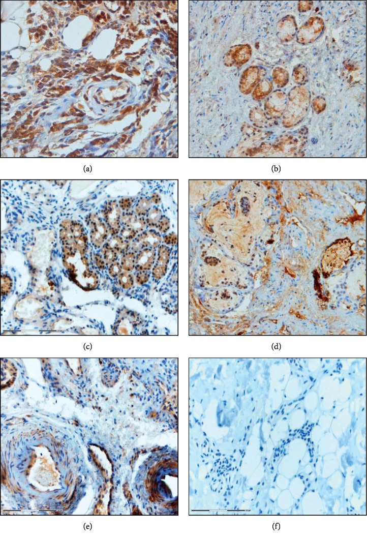
The body's autoimmune process is involved in the development of Graves' disease (GD), which is manifested by an overactive thyroid gland. In some patients, autoreactive inflammatory reactions contribute to the development of symptoms such as thyroid ophthalmopathy, and the subsequent signs and symptoms are derived from the expansion of orbital adipose tissue and edema of extraocular muscles within the orbit. The autoimmune process, production of antibodies against self-antigens such as TSH receptor (TSHR) and IGF-1 receptor (IGF-1R), inflammatory infiltration, and accumulation of glycosaminoglycans (GAG) lead to edematous-infiltrative changes in periocular tissues. As a consequence, edema exophthalmos develops. Orbital fibroblasts seem to play a crucial role in orbital inflammation, tissue expansion, remodeling, and fibrosis because of their proliferative activity as well as their capacity to differentiate into adipocytes and myofibroblasts and production of GAG. In this paper, based on the available medical literature, the immunological mechanism of GO pathogenesis has been summarized. Particular attention was paid to the role of orbital fibroblasts and putative autoantigens. A deeper understanding of the pathomechanism of the disease and the involvement of immunological processes may give rise to the introduction of new, effective, and safe methods of treatment or monitoring of the disease activity.


Conflict of interest statement

The authors declare that they have no conflicts of interest.

Figures

Figure 1
Figure 1
Pathogenesis of Graves' disease (GD) and Graves' ophthalmopathy (GO). GD is an autoimmune disease in which antibodies stimulate the thyroid to produce thyroid hormones leading to hyperthyroidism. One of the most common signs and symptoms is enlargement of the thyroid gland (goiter) while GO is the most frequent extrathyroidal involvement of GD. Inflammation and infiltration extraocular tissues result in edema and fibrosis of these tissues.
Figure 2
Figure 2
Pathogenesis of edematous-infiltrative changes. Inflammatory infiltration in periocular tissues and activity of orbital fibroblasts lead to expansion and remodeling of tissues. Increased intraocular pressure within the inflexible bony orbit results in proptosis and can contribute to developing optic nerve compression.
Figure 3
Figure 3
The proportion of T lymphocytes in the pathogenesis of Graves' ophthalmopathy. The initial phase of GO is characterized by increased activity of Th1 lymphocyte-producing cytokines that enhance fibroblast proliferation and GAG production. Th2 lymphocytes involved in the late phase participate in remodeling and fibrosis of periorbital tissues.
Figure 4
Figure 4
The participation of orbital fibroblasts in orbital inflammation. Cytokines, growth factors, and T cells stimulate orbital fibroblasts to produce chemokines and cytokines. PGE2 produced by orbital fibroblasts activates mast cells and B-cell maturation as well as stimulates the production of IL-6 by orbital fibroblasts.
Figure 5
Figure 5
The participation of orbital fibroblasts in orbital tissue remodeling. Orbital fibroblasts express TSHR, IGF-1R, and CD40. Infiltrated immune cells, antibodies, secreted cytokines, chemokines, growth factors, and also CD40-CD154 interactions activate orbital fibroblasts. Inflammatory mediators (Il-1β and IL-6) that enhance adipogenesis activate Thy1− orbital fibroblasts to differentiate into adipocytes. And Thy1+ orbital fibroblasts (with CD90 expression), activated by TGF-β, differentiate into myofibroblasts. Proliferative activity of orbital fibroblasts, their differentiation, and capacity to synthesize extracellular matrix contribute to orbital tissue expansion, remodeling, and fibrosis.
Figure 6
Figure 6
Immunohistochemistry on formalin-fixed paraffin-embedded tissue section of fat tissue of the eye socket obtained from patients who underwent endoscopic orbital decompression due to dysthyroid optic neuropathy: (a) TGF-β (×200); (b) TLR-4 (×100); (c) NF-kappa B (×100); (d) HIF-1α (×100); (e) IL-17 (×100); (f) isotype control (×100). The color reaction was visualized using DAB as a chromogen.

Similar articles

Cited by

References

    1. Smith T. J., Hegedüs L. Graves’ disease. New England Journal of Medicine. 2016;375(16):1552–1565. doi: 10.1056/nejmra1510030. - DOI - PubMed
    1. Piantanida E. Preoperative management in patients with Graves’ disease. Gland Surgery. 2017;6(5):476–481. - PMC - PubMed
    1. Lin J. D., Yang S. F., Wang Y. H., et al. Associations of melatonin receptor gene polymorphisms with Graves’ disease. PLoS One. 2017;12(9) doi: 10.1371/journal.pone.0185529.e0185529 - DOI - PMC - PubMed
    1. Khong J. J., McNab A. A., Ebeling P. R., Craig J. E., Selva D. Pathogenesis of thyroid eye disease: review and update on molecular mechanisms. British Journal of Ophthalmology. 2016;100(1):142–150. doi: 10.1136/bjophthalmol-2015-307399. - DOI - PubMed
    1. Salvi M., Vannucchi G., Currò N., et al. Efficacy of B-cell targeted therapy with rituximab in patients with active moderate to severe Graves’ orbitopathy: a randomized controlled study. The Journal of Clinical Endocrinology & Metabolism. 2015;100(2):422–431. doi: 10.1210/jc.2014-3014. - DOI - PMC - PubMed