Review
. 2020 Jun;77(11):2029-2048.
doi: 10.1007/s00018-019-03389-7. Epub 2019 Nov 28.

Sperm defects in primary ciliary dyskinesia and related causes of male infertility

Affiliations
Review

Sperm defects in primary ciliary dyskinesia and related causes of male infertility

Anu Sironen et al. Cell Mol Life Sci. 2020 Jun.

Abstract

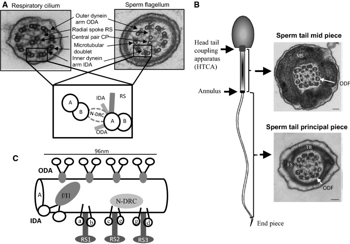
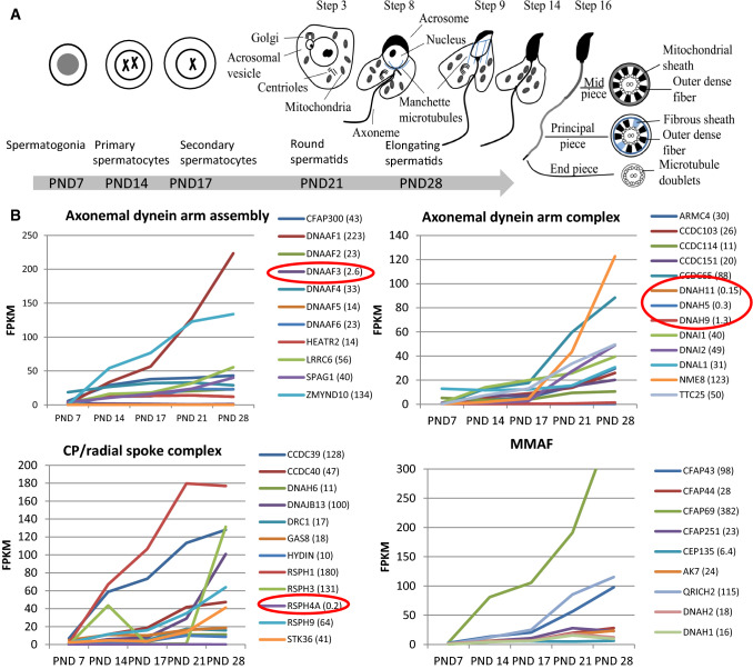
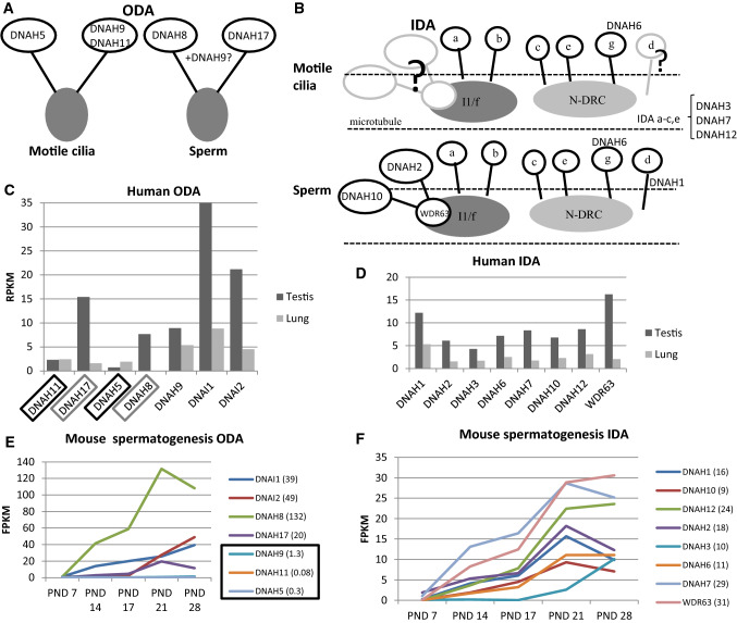
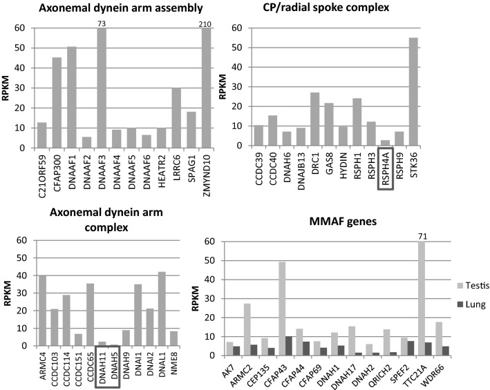
The core axoneme structure of both the motile cilium and sperm tail has the same ultrastructural 9 + 2 microtubular arrangement. Thus, it can be expected that genetic defects in motile cilia also have an effect on sperm tail formation. However, recent studies in human patients, animal models and model organisms have indicated that there are differences in components of specific structures within the cilia and sperm tail axonemes. Primary ciliary dyskinesia (PCD) is a genetic disease with symptoms caused by malfunction of motile cilia such as chronic nasal discharge, ear, nose and chest infections and pulmonary disease (bronchiectasis). Half of the patients also have situs inversus and in many cases male infertility has been reported. PCD genes have a role in motile cilia biogenesis, structure and function. To date mutations in over 40 genes have been identified cause PCD, but the exact effect of these mutations on spermatogenesis is poorly understood. Furthermore, mutations in several additional axonemal genes have recently been identified to cause a sperm-specific phenotype, termed multiple morphological abnormalities of the sperm flagella (MMAF). In this review, we discuss the association of PCD genes and other axonemal genes with male infertility, drawing particular attention to possible differences between their functions in motile cilia and sperm tails.

Keywords: Axoneme; Cilia; Dynein; Infertility; MMAF; Motility; PCD; Sperm tail.


Figures

Fig. 1
Fig. 1
Axonemal structure of respiratory cilia and sperm flagella. a The shared electron microscopic axonemal structure of the 9 + 2 motile respiratory cilia and sperm flagellum with major structural features indicated. Outer doublet microtubules are connected to the central pair (CP) by radial spokes (RS) and to each other by nexin links (nexin–dynein regulatory complex, N-DRC). Outer and inner dynein arms (ODA and IDA, respectively) provide the energy for the movement. b Accessory structures of the sperm tail. The 9 + 2 axoneme is surrounded by outer dense fibres (ODF) in the mid and principal piece of the sperm tail. Along the mid piece the ODFs are surrounded by mitochondria and along the principal piece the fibrous sheath replaces ODFs 3 and 8 and the transverse ribs (TR) encircle ODFs. The mid-piece and principal piece are separated by the annulus and the tail is connected to the head by the head tail coupling apparatus (HTCA). The formation of sperm tail structures and associated proteins have been previously reviewed [58]. c Schematic presentation of the protein complex 96 nm repeat units along the A doublet microtubule. The complex is formed of four identical ODAs, one two-headed IDA (f/I1), six single-headed IDAs (a–e and g), three RS and a single N-DRC
Fig. 2
Fig. 2
PCD gene expression during the first wave of mouse spermatogenesis. a Cell type appearance of mouse sperm during the first wave of spermatogenesis. In addition to specific populations of male germ cells, all samples contain somatic cells. PND post-natal day. b PCD gene expression changes during the progression of mouse spermatogenesis, divided up according to the different functional categories of PCD genes. RNAseq was conducted on testis tissue samples collected at specific postnatal time points as indicated in 3A [56]. Red circles indicate the detected levels of genes Dnah5, Dnah9, Dnah11 and Rsph4a, showing their very low levels of expression as discussed in the text, which supports a potentially lesser role in sperm compared to their known motile cilia-specific functions. In the mouse testis, Dnaaf3 expression was also limited. All identified MMAF genes show a pattern of increasing expression during the progression of spermatogenesis. The highest reads per kilobase million (RPKM) expression value is indicated in brackets
Fig. 3
Fig. 3
Expression of axonemal dynein arm genes in motile cilia and during spermatogenesis. A model of suggested differences in components of ODA (a) and IDA (b) in motile cilia versus sperm, based on evidence of expression studies from Drosophila [128], human [26] and mouse [50]. The IDA is a suggested model, because in humans there are approximately 7 isoforms [1 double headed dynein (I1/f) and 6 single dynein IDA complexes (a–e, g)] which are thought to be structurally different within an axonemal 96-nm repeating pattern [104]. The human IDA structure is much less resolved in terms of its possible subunit components and more complicated than the human ODA. ODA (c) and IDA (d) DNAH gene expression in the human testis and lung [26]. ODA component DNAH8 is not expressed in the lung and DNAH17 shows low expression (grey box). In contrast, DNAH5 and DNAH11 have low expression in the testis (black box) compared to the lung. DNAH2, DNAH10 and DNAH binding gene WDR63 show low transcript levels in the lung, but DNAH1 expression in relatively high compared to axonemal gene expression in general. RPKM reads per kilobase million. ODA (e) and IDA (f) gene expression during the first wave of mouse spermatogenesis (see Fig. 2 for graph of cell content during the first wave of spermatogenesis in mouse). Dnah5, Dnah9 and Dnah11 show very low expression during mouse spermatogenesis [black box, fragments per kilobase million (FPKM) expression levels indicated in brackets]. All IDA genes show high expression at PND21 during the axoneme formation, except Dnah3 may be required later during the spermatid elongation
Fig. 4
Fig. 4
PCD gene expression in the human testis and MMAF gene expression in the lung. PCD genes are divided up into functional categories in each graph. DNAH5, DNAH11 and RSPH4A show markedly low expression (RPKM < 3) in the testis (boxes) compared to other PCD genes. The high RPKM value for DNAAF3, ZMYND10 and TTC21A is indicated above the bar (73, 210 and 71, respectively). Some expression is also detected for MMAF genes in the lung. The data was produced by RNAseq of human tissues samples [29]

References

    1. Abbasi F, Miyata H, Shimada K, Morohoshi A, Nozawa K, Matsumura T, Xu Z, Pratiwi P, Ikawa M. RSPH6A is required for sperm flagellum formation and male fertility in mice. J Cell Sci. 2018 doi: 10.1242/jcs.221648. - DOI - PMC - PubMed
    1. Amiri-Yekta A, Coutton C, Kherraf ZE, Karaouzene T, Le Tanno P, Sanati MH, Sabbaghian M, Almadani N, Sadighi Gilani MA, Hosseini SH, Bahrami S, Daneshipour A, Bini M, Arnoult C, Colombo R, Gourabi H, Ray PF. Whole-exome sequencing of familial cases of multiple morphological abnormalities of the sperm flagella (MMAF) reveals new DNAH1 mutations. Hum Reprod. 2016;31:2872–2880. doi: 10.1093/humrep/dew262. - DOI - PubMed
    1. Antony D, Becker-Heck A, Zariwala MA, Schmidts M, Onoufriadis A, Forouhan M, Wilson R, Taylor-Cox T, Dewar A, Jackson C, Goggin P, Loges NT, Olbrich H, Jaspers M, Jorissen M, Leigh MW, Wolf WE, Daniels ML, Noone PG, Ferkol TW, Sagel SD, Rosenfeld M, Rutman A, Dixit A, O’Callaghan C, Lucas JS, Hogg C, Scambler PJ, Emes RD, Uk10k. Chung EM, Shoemark A, Knowles MR, Omran H, Mitchison HM. Mutations in CCDC39 and CCDC40 are the major cause of primary ciliary dyskinesia with axonemal disorganization and absent inner dynein arms. Hum Mutat. 2013;34:462–472. doi: 10.1002/humu.22261. - DOI - PMC - PubMed
    1. Auguste Y, Delague V, Desvignes JP, Longepied G, Gnisci A, Besnier P, Levy N, Beroud C, Megarbane A, Metzler-Guillemain C, Mitchell MJ. Loss of calmodulin- and radial-spoke-associated complex protein CFAP251 leads to immotile spermatozoa lacking mitochondria and infertility in men. Am J Hum Genet. 2018;103:413–420. doi: 10.1016/j.ajhg.2018.07.013. - DOI - PMC - PubMed
    1. Baccetti B, Collodel G, Estenoz M, Manca D, Moretti E, Piomboni P. Gene deletions in an infertile man with sperm fibrous sheath dysplasia. Hum Reprod. 2005;20:2790–2794. doi: 10.1093/humrep/dei126. - DOI - PubMed