The wmN1 Enhancer Region of the Mouse Myelin Proteolipid Protein Gene (mPlp1) is Indispensable for Expression of an mPlp1-lacZ Transgene in Both the CNS and PNS
- PMID: 31782102
- PMCID: PMC7060809
- DOI: 10.1007/s11064-019-02919-w
The wmN1 Enhancer Region of the Mouse Myelin Proteolipid Protein Gene (mPlp1) is Indispensable for Expression of an mPlp1-lacZ Transgene in Both the CNS and PNS
Abstract
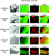
The myelin proteolipid protein gene (PLP1) encodes the most abundant protein in CNS myelin. Expression of the gene must be strictly regulated, as evidenced by human X-linked leukodystrophies resulting from variations in PLP1 copy number, including elevated dosages as well as deletions. Recently, we showed that the wmN1 region in human PLP1 (hPLP1) intron 1 is required to promote high levels of an hPLP1-lacZ transgene in mice, using a Cre-lox approach. The current study tests whether loss of the wmN1 region from a related transgene containing mouse Plp1 (mPlp1) DNA produces similar results. In addition, we investigated the effects of loss of another region (ASE) in mPlp1 intron 1. Previous studies have shown that the ASE is required to promote high levels of mPlp1-lacZ expression by transfection analysis, but had no effect when removed from the native gene in mouse. Whether this is due to compensation by another regulatory element in mPlp1 that was not included in the mPlp1-lacZ constructs, or to differences in methodology, is unclear. Two transgenic mouse lines were generated that harbor mPLP(+)Z/FL. The parental transgene utilizes mPlp1 sequences (proximal 2.3 kb of 5'-flanking DNA to the first 37 bp of exon 2) to drive expression of a lacZ reporter cassette. Here we demonstrate that mPLP(+)Z/FL is expressed in oligodendrocytes, oligodendrocyte precursor cells, olfactory ensheathing cells and neurons in brain, and Schwann cells in sciatic nerve. Loss of the wmN1 region from the parental transgene abolished expression, whereas removal of the ASE had no effect.
Keywords: Cre/loxP and Flp/Frt recombination; Enhancer; Gene regulation; Myelin proteolipid protein gene (Plp1); Transgenic mouse; lacZ.
Conflict of interest statement
Conflict of Interest
The authors declare that there are no conflicts of interest.
Figures


Similar articles
-
The wmN1 enhancer region in intron 1 is required for expression of human PLP1.Glia. 2018 Aug;66(8):1763-1774. doi: 10.1002/glia.23339. Epub 2018 Apr 23. Glia. 2018. PMID: 29683207 Free PMC article.
-
Control of human PLP1 expression through transcriptional regulatory elements and alternatively spliced exons in intron 1.ASN Neuro. 2015 Feb 18;7(1):1759091415569910. doi: 10.1177/1759091415569910. Print 2015 Jan-Feb. ASN Neuro. 2015. PMID: 25694552 Free PMC article.
-
Targeted deletion of the antisilencer/enhancer (ASE) element from intron 1 of the myelin proteolipid protein gene (Plp1) in mouse reveals that the element is dispensable for Plp1 expression in brain during development and remyelination.J Neurochem. 2013 Feb;124(4):454-65. doi: 10.1111/jnc.12092. Epub 2012 Dec 21. J Neurochem. 2013. PMID: 23157328 Free PMC article.
-
Effects of Intron 1 Sequences on Human PLP1 Expression: Implications for PLP1-Related Disorders.ASN Neuro. 2017 Jul-Aug;9(4):1759091417720583. doi: 10.1177/1759091417720583. ASN Neuro. 2017. PMID: 28735559 Free PMC article. Review.
-
The myelin proteolipid DMα in fishes.Neuron Glia Biol. 2010 May;6(2):109-12. doi: 10.1017/S1740925X09000131. Epub 2009 Jun 10. Neuron Glia Biol. 2010. PMID: 19508742 Review.
Cited by
-
PLP1-lacZ transgenic mice reveal that splice variants containing "human-specific" exons are relatively minor in comparison to the archetypal transcript and that an upstream regulatory element bolsters expression during early postnatal brain development.Front Cell Neurosci. 2023 Jan 11;16:1087145. doi: 10.3389/fncel.2022.1087145. eCollection 2022. Front Cell Neurosci. 2023. PMID: 36713780 Free PMC article.
-
Plp1 in the enteric nervous system is preferentially expressed during early postnatal development in mouse as DM20, whose expression appears reliant on an intronic enhancer.Front Cell Neurosci. 2023 May 24;17:1175614. doi: 10.3389/fncel.2023.1175614. eCollection 2023. Front Cell Neurosci. 2023. PMID: 37293625 Free PMC article.
References
-
- Macklin WB, Campagnoni CW, Deininger PL, Gardinier MV (1987) Structure and expression of the mouse myelin proteolipid protein gene. J Neurosci Res 18:383–394 - PubMed
-
- Ikenaka K, Furuichi T, Iwasaki Y, Moriguchi A, Okano H, Mikoshiba K (1988) Myelin proteolipid protein gene structure and its regulation of expression in normal and jimpy mutant mice. J Mol Biol 199:587–596 - PubMed
-
- Wight PA, Dobretsova A (1997) The first intron of the myelin proteolipid protein gene confers cell type-specific expression by a transcriptional repression mechanism in non-expressing cell types. Gene 201:111–117 - PubMed
MeSH terms
Substances
Grants and funding
LinkOut - more resources
Full Text Sources

