Review
. 2020 Apr;32(13):e1903367.
doi: 10.1002/adma.201903367. Epub 2019 Nov 29.

Biomaterials as Tools to Decode Immunity

Affiliations
Review

Biomaterials as Tools to Decode Immunity

Haleigh B Eppler et al. Adv Mater. 2020 Apr.

Abstract

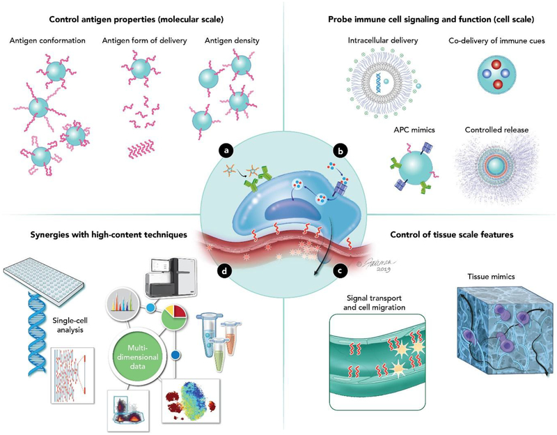
The immune system has remarkable capabilities to combat disease with exquisite selectivity. This feature has enabled vaccines that provide protection for decades and, more recently, advances in immunotherapies that can cure some cancers. Greater control over how immune signals are presented, delivered, and processed will help drive even more powerful options that are also safe. Such advances will be underpinned by new tools that probe how immune signals are integrated by immune cells and tissues. Biomaterials are valuable resources to support this goal, offering robust, tunable properties. The growing role of biomaterials as tools to dissect immune function in fundamental and translational contexts is highlighted. These technologies can serve as tools to understand the immune system across molecular, cellular, and tissue length scales. A common theme is exploiting biomaterial features to rationally direct how specific immune cells or organs encounter a signal. This precision strategy, enabled by distinct material properties, allows isolation of immunological parameters or processes in a way that is challenging with conventional approaches. The utility of these capabilities is demonstrated through examples in vaccines for infectious disease and cancer immunotherapy, as well as settings of immune regulation that include autoimmunity and transplantation.

Keywords: immunology; immunotherapy; microparticles; nanoparticles; organ-on-a-chip; organoids.


Figures

Figure 1.
Figure 1.. Overview of how biomaterials serve as tools to decipher immune function.
a. Materials can be used to control the molecular features of antigen display (molecular scale) b. Different biomaterial properties can be exploited to study how immune cells respond to different types of stimulus (cellular scale) c. Immune cells interact with the surrounding tissue to develop particular functions (tissue scale). Two areas that materials have been useful in are studying how immune cells respond to physical forces and environmental signal molecules. d. Biomaterials can interface with new high content data techniques to more deeply probe immune function.
Figure 2.
Figure 2.. Biomaterials can control antigen conformation, form of delivery, and display density.
a. Trimer proteins self-assemble as on the surface of a virus in the native orientation. Reproduced with permission.[57] Copyright 2019, Elsevier. b. These capsids can be loaded onto ICMVs in a random fashion without NTA-functionalized lipids, or in a controlled fashion by using NTA-functionalized lipids at the base of the construct. Reproduced with permission.[57]Copyright 2019, Elsevier. c. The amount of fluorescence signal generated by fluorescently labeled antibodies that were incubated with antigen loaded ICMVs. The x-axis indicates the clone of the antibody the ICMV was treated with. Different antibody clones are reactive against different features of the antigen. This figure demonstrates that directed antigen orientation causes changes in antibody binding capability. Reproduced with permission.[57] Copyright 2019, Elsevier. d. A computational model of the self-assembled 60-mer eOD HIV antigen NP structure (left) or the MD39–8-mer of the self-assembled HIV antigen NP structure (right). The blue color depicts glycans. The green color depicts antigen (eOD – left, MD39 – right). The red color is the material core that facilitates self-assembly. These constructs have distinct physical structures which can impact the development of the immune response. Reproduced with permissions.[58] Copyright 2018, American Association for the Advancement of Science. e. QDs can be used as a platform to control the molecular density of antigen displayed to APCs. The green sphere depicts the core/shell of the quantum dots. The loaded molecule is a myelin antigen. Adapted with permissions.[66] Copyright 2017, Wiley-VCH. f. QDs with low densities of myelin antigen significantly reduced disease-induced paralysis as measured using a disease severity scale in a mouse model of MS. Reproduced with permissions.[66] Copyright 2017, Wiley-VCH. g. Increasing the density of pMHC on NPs revealed a range in which valency causes an exponential increase in the amount of an inflammatory cytokine (IFNγ) produced by T cells. Reproduced with permissions.[67] Copyright 2017, Springer Nature. h. The functional fate of cells encountering pMHC NPs is controlled in part by the pMHC density, which, above a threshold density, induce microclustering of the NPs on the T cell membrane as indicated with red arrows. Reproduced with permissions.[67] Copyright 2017, Springer Nature.
Figure 3.
Figure 3.. Biomaterials can be used to study immune cell signaling and processing by exploiting material features to guide interactions with cells.
a. A schematic depicting the design of PMs. PMs were first synthesized without any cargo. Then, miR antagonist and Poly(I:C) were added to the formulation. Finally, a model antigen OVA incorporated into these structures. Reproduced with permissions.[79] Copyright 2016, American Association of Immunologists, Inc. b. The cellular uptake of miR148ai (red) and OVA (green) are shown four hours post treatment using confocal microscopy. In these images, cells were treated with either soluble miR-148ai, soluble OVA, polypeptide micelles encapsulating miR-148ai, and polypeptide micelles with OVA and miR-148ai. These images demonstrate that only the cells treated with polypeptide micelles with OVA and miR-148ai have both signals present in the cytoplasm. Reproduced with permissions.[79] Copyright 2016, American Association of Immunologists, Inc. c. Uptake was quantified at 0.5 hours post treatment using flow cytometry. This panel indicates miR-148ai-positive cells as a measure of median fluorescence intensity. Reproduced with permissions.[79] Copyright 2016, American Association of Immunologists, Inc. d. As in panel c, uptake was quantified at 0.5 hours post treatment using flow cytometry to measure the median fluorescently intensity of fluorescently-labeled OVA in live cells. Reproduced with permissions.[79] Copyright 2016, American Association of Immunologists, Inc. e. The antigen, p(Man-TLR7), and biproduct molecules that are released in the endosome as the delivered molecule is degraded by the immune cell. Reproduced with permissions.[84] Copyright 2019, Springer Nature. f. Commercially available TLR7 agonists(gray, pink, and purple) activated DC less than the synthesized p(Man-TLR7) agonist (red). Reproduced with permissions.[84] Copyright 2019, Springer Nature. g. A schematic of the cell-based assay and representative images of DCs colocalized with particles. Bone marrow derived DCs (red – rhodamine phalloidin; blue - nucleus) were cultured with PLGA particles (green). These DCs are cultured with particles then analyzed for changes in surface protein expression or soluble cytokine secretion. Reproduced with permissions.[94] Copyright 2017, Royal Society of Chemistry. h. A schematic showing the formation of artificial neutrophils. Enzymes were embedded in an organic Zn framework. These frameworks were then encapsulated in cell membranes isolated from activated neutrophils. Reproduced with permissions.[106] Copyright 2019, Wiley-VCH. i. Artificial neutrophils generated inflammatory molecules more quickly than neutrophils isolated from mice with a model of cancer. Reproduced with permissions.[106] Copyright, Wiley-VCH.
Figure 4.
Figure 4.. Biomaterials can be used to study how tissue level processes impact immunological outcomes.
a. A schematic of the microfluidics device to deliver signals to different tissue locations. The green port indicates the controlled delivery of stimulus through a specific port to the tissue slice located above. Reproduced with permissions.[111] Copyright 2017, Royal Society of Chemistry. b. These are image depicting the local stimulation of LN slices labeled with Alexa Fluor 647-glucose-BSA (red), nucleus (blue), and B cell zone (green). White arrows point to the site at which signal was delivered. In the top images, fluorescent glucose was delivered to the T cell zone of the LN. In the bottom images, fluorescent glucose was delivered to the B cell zone. Reproduced with permissions.[111] Copyright 2017, Royal Society of Chemistry. c. Quantification of images in b. Reproduced with permissions.[111] Copyright 2017, Royal Society of Chemistry. d. A schematic demonstrating the synthesis of silicate NPs encapsulated within a gelatin hydrogel. Adapted with permissions.[113] Copyright 2015, Elsevier. e. A schematic of how the 3D B cell follicle organoids are formed. These organoids contain primary B cells isolated from spleens, cultured with 40LB stromal cells and soluble signals to support growth into the hydrogel depicted in panel c. Adapted with permissions.[114] Copyright 2017, Springer Nature. f. By altering the percent weight by volume of PEG diacrylate it was possible to tune the hydrogel stiffness as measured by elastic modulus. Reproduced with permissions. [27] Copyright 2019, Wiley-VCH. g. T cells proliferated more with less stiff hydrogels as measured by a lower elastic modulus. Reproduced with permissions.[27] Copyright 2019, Wiley-VCH. h. Collagen (coll) hydrogels, collagen hydrogels functionalized with hyaluronan (HA), or functionalized with sulfate hyaluronan (sHA) were created. Hydrogels were crosslinked with EDC had greater stiffness than the other formulations. A higher IL10:IL12 ratio indicates immune cells with more regulatory functions. This figure demonstrates two-dimensional and three-dimensional culture systems differentially impact immune cell function. Reproduced with permissions.[116] Copyright 2017, Wiley-VCH.
Figure 5.
Figure 5.. Coupling biomaterials with new high content data and cellular analysis techniques creates synergistic opportunities.
a. A schematic depicting how droplets are formed that encapsulate T cells and cancer cells. Briefly, T cells and cancer cells flow alongside each other before passing through oil that creates a droplet containing one cell of each type. Reproduced with permissions.[142] Copyright 2018, Royal Society of Chemistry. b. Cell containing droplets can be sorted on the basis of cellular processes such as T cell activation. This process allows for more detailed downstream analysis of cells with characteristics of interest. Reproduced with permissions.[142] Copyright 2018, Royal Society of Chemistry. c. A schematic depicting how droplet encapsulation can be combined with single cell RNA sequencing. This allows for cells to be coencapsulated with unique RNA molecules that allow for separation of RNA from each cell.[147] d. Single cell RNA sequencing was able to differentiate between the types of cells the RNA was isolated from in pure (left) or mixed (right) cultures. Reproduced with permissions.[147] Copyright 2017, Springer Nature. e. A schematic depicting microneedles that are capable of sampling the fluid and cells that are present in the interstitial space and epidermis. These microneedles are covered in a hydrogel layer that swells in interstitial fluid and capture infiltrating immune cells. Reproduced with permissions.[149] Copyright 2018, American Association for the Advancement of Science. f. Following immunization, the percent of antigen specific cells detected using sampling microneedles was the same as in traditional blood measurements. This demonstrates that microneedles have the ability to capture the same trends as traditional sampling methods such as blood draws. Reproduced with permissions.[149] Copyright 2018, American Association for the Advancement of Science.

Similar articles

Cited by

References

    1. Ehreth J, Vaccine 2003, 21, 596. - PubMed
    1. Flasche S, Jit M, Rodríguez-Barraquer I, Coudeville L, Recker M, Koelle K, Milne G, Hladish TJ, Perkins TA, Cummings DAT, Dorigatti I, Laydon DJ, España G, Kelso J, Longini I, Lourenco J, Pearson CAB, Reiner RC, Mier-y-Terán-Romero L, Vannice K, Ferguson N, PLoS Med 2016, 13, 1. - PMC - PubMed
    1. Obando-Pacheco P, Rivero-Calle I, Gómez-Rial J, Rodríguez-Tenreiro Sánchez C, Martinón-Torres F, Vaccine 2018, 36, 5485. - PubMed
    1. Paules CI, Marston HD, Fauci AS, Engl N. J. Med 2019, NEJMp1905099. - PubMed
    1. Draper E, Bissett SL, Howell-Jones R, Waight P, Soldan K, Jit M, Andrews N, Miller E, Beddows S, PLoS One 2013, 8, DOI 10.1371/journal.pone.0061825. - DOI - PMC - PubMed

LinkOut - more resources