Review
. 2020 Feb;190(2):272-285.
doi: 10.1016/j.ajpath.2019.10.012. Epub 2019 Nov 26.

Necroptosis in the Pathophysiology of Disease

Affiliations
Review

Necroptosis in the Pathophysiology of Disease

Mitri K Khoury et al. Am J Pathol. 2020 Feb.

Abstract

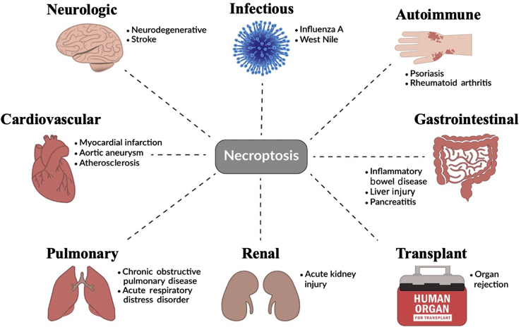
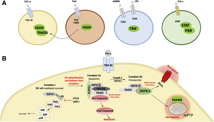
Over the past 15 years, elegant studies have demonstrated that in certain conditions, programed cell death resembles necrosis and depends on a unique molecular pathway with no overlap with apoptosis. This form of regulated necrosis is represented by necroptosis, in which the receptor-interacting protein kinase-3 and its substrate mixed-lineage kinase domain-like protein play a crucial role. With the development of knockout mouse models and molecular inhibitors unique to necroptotic proteins, this cell death has been found to occur in virtually all tissues and diseases evaluated. There are different immunologic consequences depending on whether cells die through apoptosis or necroptosis. Therefore, distinguishing between these two forms of cell death may be crucial during pathologic evaluations. In this review, we provide an understanding of necroptotic cell-death and highlight diseases in which necroptosis has been found to play a role. We also discuss the inhibitors of necroptosis and the ways these inhibitors have been used in preclinical models of diseases. These two discussions offer an understanding of the role of necroptosis in diseases and will foster efforts to pharmacologically target this unique yet pervasive form of programed cell death in the clinic.


Figures

Figure 1
Figure 1
Necroptosis plays a role in the pathogenesis of various diseases across the body, including conditions of the neurologic, cardiovascular, pulmonary, and gastrointestinal systems. Necroptosis also plays a role in infectious and autoimmune diseases. Recently, necroptosis was reported to mediate organ rejection in both cardiac and renal allografts.,
Figure 2
Figure 2
Signal transduction events downstream of tumor necrosis factor receptor 1 (TNF-RI) that cause necroptosis. A: Overall schematic highlighting the unique receptors and intracellular signal-transduction components that activate necroptosis on binding to their ligands. The receptors include TNF-receptor superfamily (TNF-RI and Fas/CD95), Toll-like-receptor superfamily (TLR3/4), and interferon receptor (IFNR). The signal transducers are in green circles. Note: all signaling components converge on RIP3 for the execution of necroptosis and an example of the downstream events is shown in B. B: Signal transduction events downstream of TNF-RI that cause necroptosis. Activation of the TNF-RI, by engagement of TNF-α, can trigger the formation of a prosurvival complex (Complex I), which contains receptor-interacting protein kinase-1 (RIPK1). When Complex I is ubiquitinated, this leads to NF-κB mediated survival. Alternately, de-ubiquitination of RIPK1 by either ubiquitin carboxyl-terminal hydolase (CYLD) or pharmacologic targeting of cellular inhibitors of apoptosis (cIAPs) can activate complex IIa. Complex IIa is protein ensemble consisting of tumor necrosis factor receptor type 1 associated death domain protein (TRADD), Fas-associated protein with death domain (FADD), and RIP1. In the presence of caspase 8, complex II preferentially drives toward IIa leading to apoptosis. However, in the absence of caspase 8 and presence of RIP3, complex II switches to IIb, which is pronecroptotic. Complex IIb then leads to necroptosis via the phosphorylation of mixed-lineage kinase domain–like pseudokinase (MLKL) by RIPK3 or its association of phosphoglycerate mutase family member (PGAM)-5 with RIPK3 that causes opening of the mitochondrial permeability transition pore complexes (mPTPs). CaMKII, Ca2+/calmodulin-dependent protein kinase II; FLIP, FLICE-like inhibitory protein; JNK, c-Jun N-terminal kinase; LPS, lipopolysaccharide; PKR, protein kinase R; RHIM, RIPK homotypic interaction motif; TLR, Toll-like receptor; TRIF, TIR-domain-containing adapter-inducing interferon-β.

References

    1. Lau A., Wang S., Jiang J., Haig A., Pavlosky A., Linkermann A., Zhang Z.X., Jevnikar A.M. RIPK3-mediated necroptosis promotes donor kidney inflammatory injury and reduces allograft survival. Am J Transplant. 2013;13:2805–2818. - PubMed
    1. Kwok C., Pavlosky A., Lian D., Jiang J., Huang X., Yin Z., Liu W., Haig A., Jevnikar A.M., Zhang Z.X. Necroptosis is involved in CD4+T cell-mediated microvascular endothelial cell death and chronic cardiac allograft rejection. Transplantation. 2017;101:2026–2037. - PubMed
    1. Gupta K., Phan N., Wang Q., Liu B. Necroptosis in cardiovascular disease - a new therapeutic target. J Mol Cell Cardiol. 2018;118:26–35. - PMC - PubMed
    1. Lin Y., Devin A., Rodriguez Y., Liu Z.G. Cleavage of the death domain kinase RIP by caspase-8 prompts TNF-induced apoptosis. Genes Dev. 1999;13:2514–2526. - PMC - PubMed
    1. Feng S., Yang Y., Mei Y., Ma L., Zhu D.E., Hoti N., Castanares M., Wu M. Cleavage of RIP3 inactivates its caspase-independent apoptosis pathway by removal of kinase domain. Cell Signal. 2007;19:2056–2067. - PubMed

Publication types