Review
. 2019 Nov 27;11(12):1887.
doi: 10.3390/cancers11121887.

Photoimmunotherapy of Ovarian Cancer: A Unique Niche in the Management of Advanced Disease

Affiliations
Review

Photoimmunotherapy of Ovarian Cancer: A Unique Niche in the Management of Advanced Disease

Shubhankar Nath et al. Cancers (Basel). .

Abstract

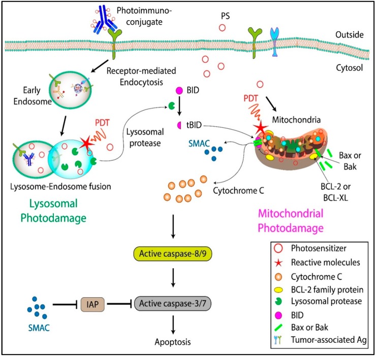
Ovarian cancer (OvCa) is the leading cause of gynecological cancer-related deaths in the United States, with five-year survival rates of 15-20% for stage III cancers and 5% for stage IV cancers. The standard of care for advanced OvCa involves surgical debulking of disseminated disease in the peritoneum followed by chemotherapy. Despite advances in treatment efficacy, the prognosis for advanced stage OvCa patients remains poor and the emergence of chemoresistant disease localized to the peritoneum is the primary cause of death. Therefore, a complementary modality that is agnostic to typical chemo- and radio-resistance mechanisms is urgently needed. Photodynamic therapy (PDT), a photochemistry-based process, is an ideal complement to standard treatments for residual disease. The confinement of the disease in the peritoneal cavity makes it amenable for regionally localized treatment with PDT. PDT involves photochemical generation of cytotoxic reactive molecular species (RMS) by non-toxic photosensitizers (PSs) following exposure to non-harmful visible light, leading to localized cell death. However, due to the complex topology of sensitive organs in the peritoneum, diffuse intra-abdominal PDT induces dose-limiting toxicities due to non-selective accumulation of PSs in both healthy and diseased tissue. In an effort to achieve selective damage to tumorous nodules, targeted PS formulations have shown promise to make PDT a feasible treatment modality in this setting. This targeted strategy involves chemical conjugation of PSs to antibodies, referred to as photoimmunoconjugates (PICs), to target OvCa specific molecular markers leading to enhanced therapeutic outcomes while reducing off-target toxicity. In light of promising results of pilot clinical studies and recent preclinical advances, this review provides the rationale and methodologies for PIC-based PDT, or photo-immunotherapy (PIT), in the context of OvCa management.

Keywords: EGFR; Ovarian cancer; photodynamic therapy; photoimmunoconjugates; photoimmunotherapy; targeted therapy.


Conflict of interest statement

The authors declare no conflicts of interest.

Figures

Figure 1
Figure 1
Photobiological and photochemical overview of photodynamic therapy (PDT). (A) Process of clinical PDT; the photosensitizer (PS) is administered systemically, followed by an appropriate PS-light interval, where the administered PS preferentially accumulates at the tumor site. Near-infrared (NIR) irradiation of the target tissue leads to localized tumor destruction. (B) Schematic representation of the Jablonski diagram showing the ground state of the PS (PS1) and the subsequent shift to a high energy excited state (PS1*) upon NIR irradiation. The PS in the excited state (PS1*) can either emit energy in the form of fluorescence radiation and relax to the ground state or undergo intersystem crossing to generate a long-lived excited triplet state (PS3*). Energy and electron transfer from the excited triplet state (PS3*) to biomolecules, water, triplet ground state oxygen, etc. leads to the formation of cytotoxic reactive molecular species (RMS). Depending on the dose of the RMS, the target tissues may either survive or undergo apoptosis/necrosis. Adapted from Nath et al. (2019) [26].
Figure 2
Figure 2
Mechanisms of cell death induced by free PS and photoimmunoconjugates (PICs). PICs bind to the corresponding receptor on the cell surface and are internalized through a receptor-mediated endocytosis pathway. The endocytosed PICs are sorted to the lysosome where they are cleaved, and the PS is released. Certain PSs diffuse through the cell membrane and localize to subcellular organelles, such as mitochondria. Depending on the PS formulation, the cells may undergo either lysosomal photodamage or mitochondrial photodamage leading to apoptosis/necrosis. Adapted from Nath et al. (2019) [26].
Figure 3
Figure 3
Comparison of predicted and actual mean survival fractions from cells treated with a combination of PIT and cisplatin (CDDP). CDDP-resistant cells were more responsive to combination treatment as compared to CDDP-sensitive cells. Adapted from Duska et al., 1999 [15].
Figure 4
Figure 4
Tumor-targeted activatable photoimmunotherapy. (A) Pictorial representation of PICs. (B) Mechanism of tumor-targeted PIC activation. (C) The PS in PICs stay quenched under normal conditions. Once internalized in the target cells, they are dequenched through lysosomal degradation for tumor-targeted activatable photoimmunotherapy (taPIT). (D) Mouse model of micrometastatic OvCa and the scheme for endoscopic fluorescence imaging. (E) Comparison of fluorescence (red) from free benzoporphyrin derivative (BPD) (upper panel) and PIC (lower panel) administered to mice in no tumor controls (left panel) and with disseminated tumors (right panel). Adapted from Spring et al. 2014 [86].
Figure 5
Figure 5
PIC design and effects. (A) Pictorial representation of direct vs indirect conjugation of the PSs to the antibody. The PS can be conjugated to the antibody either directly or through an intermediate polymer which increases the PS loading. (B) Effect of charge on cellular uptake and phototoxicity. PS conjugated to positively charged (poly-lysine conjugated) or negatively charged (succinylated poly-lysine conjugated) polymers were conjugated to the F(ab)2 fragment of OC125. The positively charged PIC led to an increase in both cellular uptake of PS and phototoxicity. Adapted from Hamblin et al. 1996 [17]. (C) Enhancing intracellular targeting by preventing lysosomal degradation. After co-incubation with PS (BPD) and fluorescein isothiocyanate-PIC (FITC-PIC), cells were sequentially irradiated with a low dose of 690 nm light to activate BPD-induced PCI and destabilize endosomal membranes, allowing PIC to be released into the cytosol. The second irradiation with 490 nm light initiates FITC-PIC mediated PIT to selectively kill Ki-67 expressing cells. Adapted from Wang et al. 2015 [123] and Wang et al. 2016 [108]. (D) Increase in BPD uptake in OvCar-5 cells due to the “carrier effect.” The binding and uptake of a single PIC results in the endocytosis of other nanoparticle-bound PICs. The “carrier effect” results in a significantly higher cellular uptake of PS and increases therapeutic outcomes in vivo. Adapted from Huang et al. 2018 [124].
Figure 6
Figure 6
Multi-functional epidermal growth factor receptor (EGFR)-targeted PIT. (A) In cancer cells, overexpressed EGFRs bind to the corresponding ligands and promote cell growth, proliferation, metastasis, angiogenesis, etc. (B) The administration of PICs targeting EGFR leads to selective accumulation of the PS in the malignant tissue and inhibition of EGFR signaling pathway and induces localized cell death upon irradiation (right panel).
Figure 7
Figure 7
(A) Mean tumor burden for mice treated with either C225 or PDT monotherapy, compared with a combination therapy of C225 and PDT. (B) Kaplan–Meier survival curves for mice treated with photodynamic therapy only, C225 only, and mice treated with a combination therapy of PDT and C225. Combination treatment with PDT and C225 resulted in a significant enhancement in survival as compared to the individual monotherapies. BPD = benzoporphyrin derivative. PDT = photodynamic therapy. C225 (Cetuximab, Anti-EGFR antibody). Adapted from del Carmen et al. 2005 [83].

References

    1. Christie E., Bowtell D. Acquired chemotherapy resistance in ovarian cancer. Ann. Oncol. 2017;28:viii13–viii15. doi: 10.1093/annonc/mdx446. - DOI - PubMed
    1. Cooke S.L., Brenton J.D. Evolution of platinum resistance in high-grade serous ovarian cancer. Lancet Oncol. 2011;12:1169–1174. doi: 10.1016/S1470-2045(11)70123-1. - DOI - PubMed
    1. Jelovac D., Armstrong D.K. Recent progress in the diagnosis and treatment of ovarian cancer. CA Cancer J. Clin. 2011;61:183–203. doi: 10.3322/caac.20113. - DOI - PMC - PubMed
    1. Cortez A.J., Tudrej P., Kujawa K.A., Lisowska K.M. Advances in ovarian cancer therapy. Cancer Chemother. Pharmacol. 2018;81:17–38. doi: 10.1007/s00280-017-3501-8. - DOI - PMC - PubMed
    1. Brackmann M., Stasenko M., Uppal S., Erba J., Reynolds R.K., McLean K. Comparison of first-line chemotherapy regimens for ovarian carcinosarcoma: A single institution case series and review of the literature. BMC Cancer. 2018;18:172. doi: 10.1186/s12885-018-4082-6. - DOI - PMC - PubMed

LinkOut - more resources