. 2019 Nov 27;8(4):268.
doi: 10.3390/pathogens8040268.

Decrease of miR-19b-3p in Brain Microvascular Endothelial Cells Attenuates Meningitic Escherichia coli-Induced Neuroinflammation via TNFAIP3-Mediated NF-κB Inhibition

Affiliations

Decrease of miR-19b-3p in Brain Microvascular Endothelial Cells Attenuates Meningitic Escherichia coli-Induced Neuroinflammation via TNFAIP3-Mediated NF-κB Inhibition

Nouman Amjad et al. Pathogens. .

Abstract

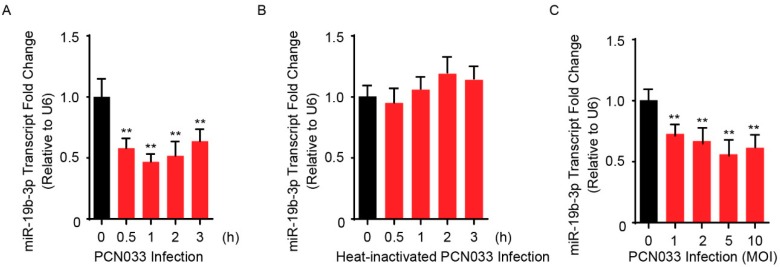
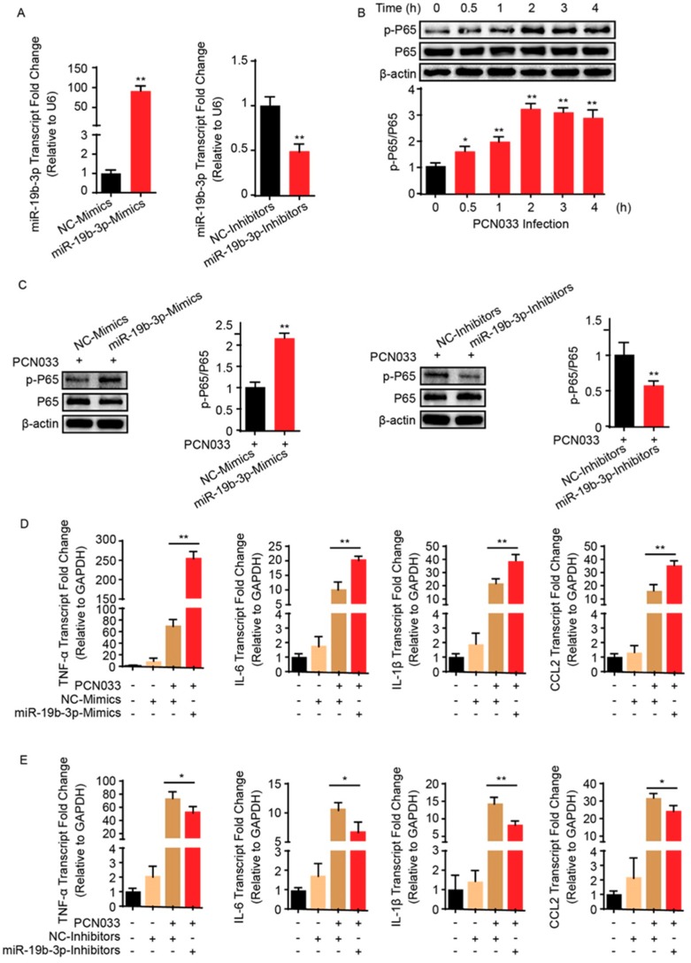
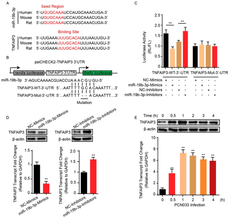
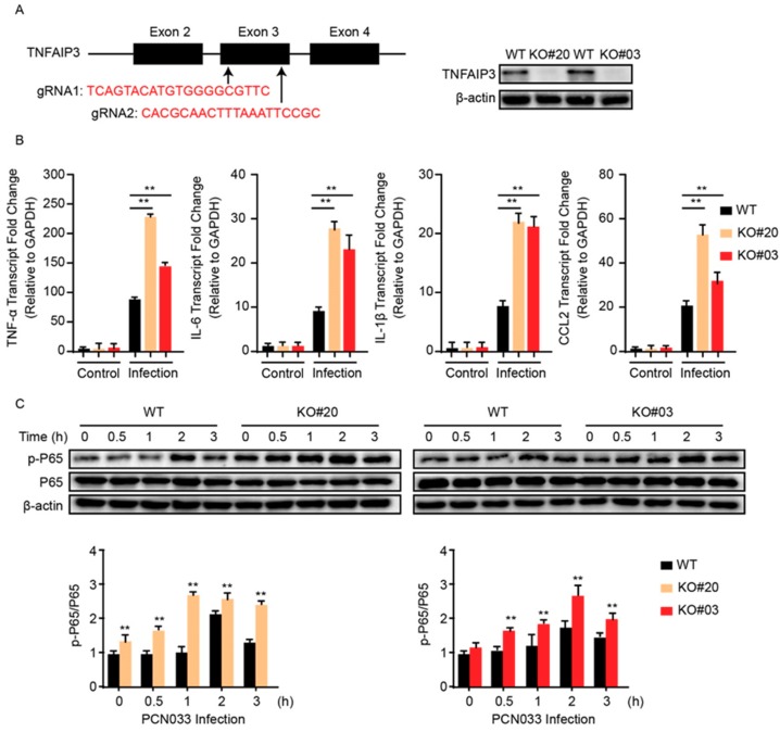
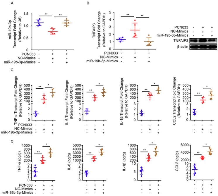
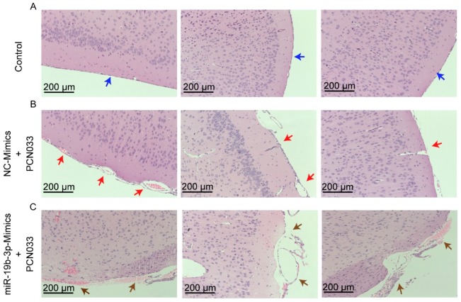
Meningitic Escherichia coli can traverse the host's blood-brain barrier (BBB) and induce severe neuroinflammatory damage to the central nervous system (CNS). During this process, the host needs to reasonably balance the battle between bacteria and brain microvascular endothelial cells (BMECs) to minimize inflammatory damage, but this quenching of neuroinflammatory responses at the BBB is unclear. MicroRNAs (miRNAs) are widely recognized as key negative regulators in many pathophysiological processes, including inflammatory responses. Our previous transcriptome sequencing revealed numbers of differential miRNAs in BMECs upon meningitic E. coli infection; we next sought to explore whether and how these miRNAs worked to modulate neuroinflammatory responses at meningitic E. coli entry of the BBB. Here, we demonstrated in vivo and in vitro that meningitic E. coli infection of BMECs significantly downregulated miR-19b-3p, which led to attenuated production of proinflammatory cytokines and chemokines via increasing the expression of TNFAIP3, a negative regulator of NF-κB signaling. Moreover, in vivo injection of miR-19b-3p mimics during meningitic E. coli challenge further aggravated the inflammatory damage to mice brains. These in vivo and in vitro findings indicate a novel quenching mechanism of the host by attenuating miR-19b-3p/TNFAIP3/NF-κB signaling in BMECs in response to meningitic E. coli, thus preventing CNS from further neuroinflammatory damage.

Keywords: NF-κB; TNFAIP3; brain microvascular endothelial cells; meningitic Escherichia coli; miR-19b-3p; neuroinflammation.


Conflict of interest statement

The authors declare no conflicts of interest.

Figures

Figure 1
Figure 1
Meningitic Escherichia coli PCN033 infection decreases the endogenous miR-19b-3p levels in human brain microvascular endothelial cells (hBMECs). (A) The significant decrease of miR-19b-3p transcription in hBMECs in response to PCN033 infection (Multiplicity of Infection, MOI = 10) at different time points. (B) Heat-inactivated PCN033 at a MOI of 10 was unable to decrease miR-19-3p levels in hBMECs at indicated time points. (C) The significant reduction of miR-19b-3p levels in hBMECs upon PCN033 infection at indicated MOI for 3 h. Data represent mean ± standard deviation (SD) from three independent experiments. U6 was used as the reference control for the miR-19b-3p quantitation. ** p < 0.01.
Figure 2
Figure 2
MiR-19b-3p enhances meningitic E. coli PCN033-induced inflammatory responses. (A) The significant increase or decrease of endogenous miR-19b-3p level in hBMECs by transfection of the synthetic miR-19b-3p mimics and inhibitors (both at final concentration of 50 nM), respectively. (B) The significant time-dependent activation of NF-κB p65 subunit in hBMECs in response to PCN033 challenge, determined by Western blot. The β-actin protein served as the loading control, and densitometry was performed to analyze the blotting bands. (C) Effects of miR-19b-3p mimic or inhibitor transfection (at 50 nM) on the phosphorylation of NF-κB p65 subunit in hBMECs in response to PCN033 infection (at Multiplicity of Infection (MOI) of 10 for 3 h). The β-actin was used as loading control, and densitometry was performed to analyze the difference among treatments. (D,E) Effects of the miR-19b-3p mimic or inhibitor transfection on the PCN033-induced production of cytokines and chemokines determined by qPCR. Data represent mean ± SD from three independent experiments. GAPDH was used as the internal reference. * p < 0.05; ** p < 0.01.
Figure 3
Figure 3
miR-19b-3p negatively regulates the expression of TNFAIP3. (A) Conservation of the miR-19b-3p sequence among different species (upper panel), and conservation of the miR-19b-3p target sequence in TNFAIP3 among different species (lower panel). Human, Homo sapiens; Mouse, Mus musculus; Rat, Rattus norvegicus. (B) The miRNA response elements (MREs) of miR-19b-3p were shown on the sequence of TNFAIP3 3’-UTR, and mutations were introduced on these MREs. Both wild-type and the mutated sequences were cloned into psiCHECK-2 plasmid. (C) 293T cells were co-transfected with miR-19b-3p mimics, miR-19b-3p inhibitors, or their corresponding control oligonucleotide (final concentration at 50 nM), together with the wild-type or mutated TNFAIP3 3’-UTR luciferase reporter plasmid, and renilla luciferase activity was measured and normalized to firefly luciferase activity after 24 h. (D) TNFAIP3 protein, as well as mRNA levels, were determined in hBMECs after 24 h of transfection with miR-19b-3p mimics, miR-19b-3p inhibitors, or their corresponding control oligonucleotide (final concentration at 50 nM). (E) Western blot and qPCR analyses of TNFAIP3 in hBMECs in response to PCN033 infection. Data represent mean ± SD from three independent experiments. Protein level was normalized to β-actin, and mRNA level as normalized to GAPDH. ** p < 0.01.
Figure 4
Figure 4
Knock-out of TNFAIP3 significantly increases meningitic E. coli-caused neuroinflammatory responses in hBMECs. (A) Schematics briefly showing the design of two gRNAs (gRNA1 and gRNA2) in the TNFAIP3 Exon 3 and the identification of TNFAIP3 knock-out (KO) cell lines KO#20 and KO#03 through Western blot analysis. (B) qPCR analysis of TNF-α, IL-6, IL-1β, and CCL2 expression in wild-type, TNFAIP KO#20, and TNFAIP KO#03 hBMECs in response to PCN033 infection. Data represent mean ± SD from three independent experiments. GAPDH was used as the internal reference. (C) Western blot analysis of the p65 phosphorylation in wild-type, TNFAIP KO#20, and TNFAIP KO#03 hBMECs in response to PCN033 infection. β-actin was used as the loading control, and densitometry was performed to analyze the differences. ** p < 0.01.
Figure 5
Figure 5
miR-19b-3p mimic pretreatment contributes to neuroinflammatory responses in meningitic E. coli-challenged mice. (A) qPCR analysis of miR-19b-3p in brain lysates from control mice, PCN033-challenged mice with or without miR-19b-3p mimic pretreatment. U6 was used as the internal reference. Data are presented as mean ± SD from five individual mice. (B) The mRNA level of TNFAIP3 in mouse brains from control mice, PCN033-challenged mice with or without miR-19b-3p mimic pretreatment was analyzed by qPCR. GAPDH was used as the internal reference. Data were presented as mean ± SD from five individual mice. The protein level of TNFAIP3 in mouse brains from control mice, PCN033-challenged mice with or without miR-19b-3p mimic pretreatment was additionally analyzed by Western blot. β-actin was used as the loading control. (C) qPCR analysis of TNF-α, IL-1β, IL-6, and CCL2 transcription from control mice, PCN033-challenged mice with or without miR-19b-3p mimic pretreatment. GAPDH was used as the internal reference. Data are presented as mean ± SD from five individual mice. (D) ECL analysis of TNF-α, IL-1β, IL-6, and CCL2 levels in brain lysates from control mice, PCN033-challenged mice with or without miR-19b-3p mimic pretreatment. Data are expressed as mean ± SD (n = 5). * p < 0.05; ** p < 0.01.
Figure 6
Figure 6
Histopathological analysis of meninges in PCN033-challenged mice with or without miR-19b-3p mimic pretreatment. (A) The normal meninges from uninfected mice (blue arrows). (B) The meninges from PCN033-challenged mice receiving the control mimics. Red arrows indicate meninges thickening, hyperemia, and inflammatory cell accumulation. (C) The meninges from PCN033-challenged mice receiving miR-19b-3p mimic pretreatment, and brown arrows show more severe thickening, hyperemia, and accumulation of inflammatory cells, as compared to the challenged brains with control mimic treatment. Scale bar = 200 μm.

References

    1. Kim K.S. Acute bacterial meningitis in infants and children. Lancet Infect. Dis. 2010;10:32–42. doi: 10.1016/S1473-3099(09)70306-8. - DOI - PubMed
    1. McGill F., Heyderman R.S., Panagiotou S., Tunkel A.R., Solomon T. Acute bacterial meningitis in adults. Lancet. 2016;388:3036–3047. doi: 10.1016/S0140-6736(16)30654-7. - DOI - PubMed
    1. Kim K.S. Pathogenesis of bacterial meningitis: From bacteraemia to neuronal injury. Nat. Rev. Neurosci. 2003;4:376–385. doi: 10.1038/nrn1103. - DOI - PubMed
    1. Coureuil M., Lecuyer H., Bourdoulous S., Nassif X. A journey into the brain: Insight into how bacterial pathogens cross blood-brain barriers. Nat. Rev. Microbiol. 2017;15:149–159. doi: 10.1038/nrmicro.2016.178. - DOI - PubMed
    1. Blanchette M., Daneman R. Formation and maintenance of the bbb. Mech. Dev. 2015;138:8–16. doi: 10.1016/j.mod.2015.07.007. - DOI - PubMed

LinkOut - more resources