

MPPa-PDT suppresses breast tumor migration/invasion by inhibiting Akt-NF-κB-dependent MMP-9 expression via ROS
- PMID: 31783821
- PMCID: PMC6884812
- DOI: 10.1186/s12885-019-6374-x
MPPa-PDT suppresses breast tumor migration/invasion by inhibiting Akt-NF-κB-dependent MMP-9 expression via ROS
Abstract
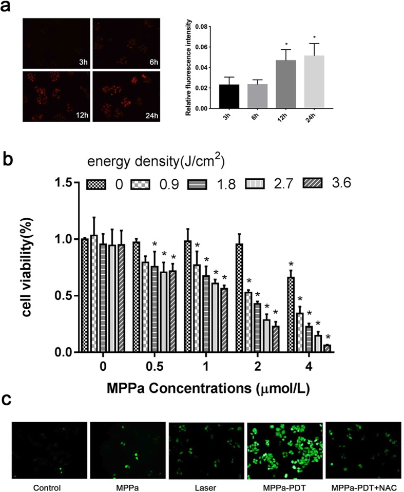
Background: Breast cancer is one of the most commonly diagnosed cancers in women, with high morbidity and mortality. Tumor metastasis is implicated in most breast cancer deaths; thus, inhibiting metastasis may provide a therapeutic direction for breast cancer. In the present study, pyropheophorbide-α methyl ester-mediated photodynamic therapy (MPPa-PDT) was used to inhibit metastasis in MCF-7 breast cancer cells.
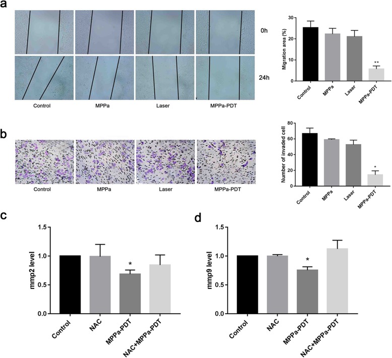
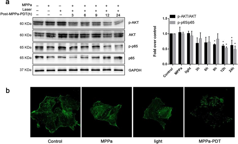
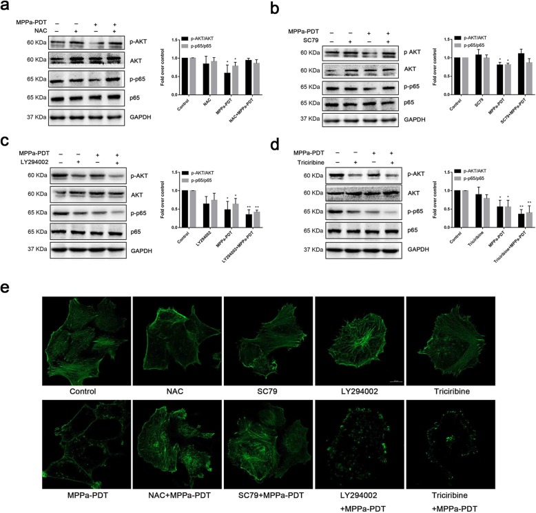
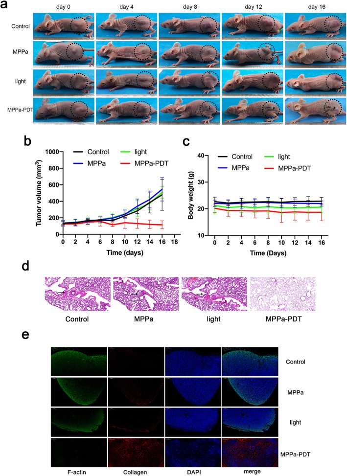
Methods: Uptake of MPPa was detected by fluorescence microscopy. Cell viability was evaluated by the Cell Counting Kit-8 (CCK-8). ROS generation was detected by 2',7'-dichlorodihydrofluorescein diacetate (DCFH-DA). The migration of cells was assessed by wound healing assay, and invasion ability was assessed by Matrigel invasion assay. Levels of MMP2 and MMP9 were measured by PCR. Akt, phospho-Akt (Ser473), phospho-NF-κB p65 (Ser536) and NF-κB p65 were measured by western blotting. The F-actin cytoskeleton was observed by immunofluorescence. Lung tissue was visualized by hematoxylin and eosin staining.
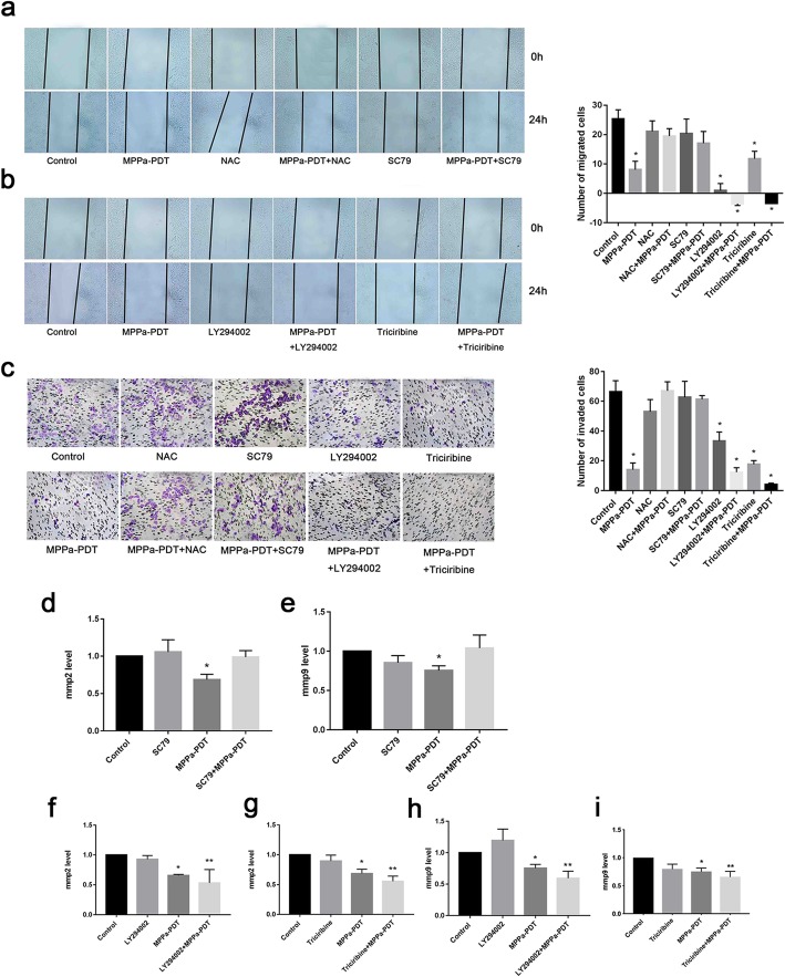
Results: Following MPPa-PDT, migration and invasion were decreased in the MCF-7 cells. MPPa-PDT downregulated the expression of MMP2 and MMP9, which are responsible for the initiation of metastasis. MPPa-PDT reduced the phosphorylation of Akt and NF-κB. MPPa-PDT also reduced the expression of F-actin in cytoskeleton in MCF-7 cells. These effects were blocked by the reactive oxygen species scavenger NAC or the Akt activator SC79, while the PI3K inhibitor LY294002 or the Akt inhibitor triciribine enhanced these effects. Moreover, MPPa-PDT inhibited tumor metastasis and destroyed F-actin in vivo.
Conclusion: Taken together, these results demonstrate that MPPa-PDT inhibits the metastasis of MCF-7 cells both in vitro and in vivo and may be involved in the Akt/NF-κB-dependent MMP-9 signaling pathway. Thus, MPPa-PDT may be a promising treatment to inhibit metastasis.
Keywords: Breast tumor; Invasion; Migration; Photodynamic therapy; Reactive oxygen species.
Conflict of interest statement
The authors declare that they have no competing interests.
Figures
Similar articles
-
Pyropheophorbide-α methyl ester-mediated photodynamic therapy induces apoptosis and inhibits LPS-induced inflammation in RAW264.7 macrophages.Photodiagnosis Photodyn Ther. 2019 Mar;25:148-156. doi: 10.1016/j.pdpdt.2018.12.002. Epub 2018 Dec 15. Photodiagnosis Photodyn Ther. 2019. PMID: 30562579
-
Curcumol Suppresses Breast Cancer Cell Metastasis by Inhibiting MMP-9 Via JNK1/2 and Akt-Dependent NF-κB Signaling Pathways.Integr Cancer Ther. 2016 Jun;15(2):216-25. doi: 10.1177/1534735416642865. Epub 2016 Apr 28. Integr Cancer Ther. 2016. PMID: 27125675 Free PMC article.
-
Establishment and characterization of human osteosarcoma cells resistant to pyropheophorbide-α methyl ester-mediated photodynamic therapy.Int J Oncol. 2017 Nov;51(5):1427-1438. doi: 10.3892/ijo.2017.4136. Epub 2017 Sep 27. Int J Oncol. 2017. PMID: 29048645 Free PMC article.
-
The role of photodynamic therapy in breast cancer - A review of in vitro research.Biomed Pharmacother. 2021 Dec;144:112342. doi: 10.1016/j.biopha.2021.112342. Epub 2021 Oct 19. Biomed Pharmacother. 2021. PMID: 34678730 Review.
-
Tumor cell survival pathways activated by photodynamic therapy: a molecular basis for pharmacological inhibition strategies.Cancer Metastasis Rev. 2015 Dec;34(4):643-90. doi: 10.1007/s10555-015-9588-7. Cancer Metastasis Rev. 2015. PMID: 26516076 Free PMC article. Review.
Cited by
-
Antitumor effects and mechanisms of pyropheophorbide‑α methyl ester‑mediated photodynamic therapy on the human osteosarcoma cell line MG‑63.Int J Mol Med. 2020 Apr;45(4):971-982. doi: 10.3892/ijmm.2020.4494. Epub 2020 Feb 10. Int J Mol Med. 2020. PMID: 32124948 Free PMC article.
-
Counteracting Cisplatin-Induced Testicular Damages by Natural Polyphenol Constituent Honokiol.Antioxidants (Basel). 2020 Aug 9;9(8):723. doi: 10.3390/antiox9080723. Antioxidants (Basel). 2020. PMID: 32784851 Free PMC article.
-
Combined Photodynamic and Photothermal Therapy and Immunotherapy for Cancer Treatment: A Review.Int J Nanomedicine. 2022 Dec 16;17:6427-6446. doi: 10.2147/IJN.S388996. eCollection 2022. Int J Nanomedicine. 2022. PMID: 36540374 Free PMC article. Review.
-
The Role of Graphene Oxide Nanocarriers in Treating Gliomas.Front Oncol. 2022 Jan 28;12:736177. doi: 10.3389/fonc.2022.736177. eCollection 2022. Front Oncol. 2022. PMID: 35155223 Free PMC article. Review.
-
Evaluation of Nanoparticles Covalently Bound with BODIPY for Their Photodynamic Therapy Applicability.Int J Mol Sci. 2024 Mar 10;25(6):3187. doi: 10.3390/ijms25063187. Int J Mol Sci. 2024. PMID: 38542161 Free PMC article.
References
-
- Lee WT, Lee TH, Cheng CH, Chen KC, Chen YC, Lin CW. Antroquinonol from antrodia camphorata suppresses breast tumor migration/invasion through inhibiting ERK-AP-1- and AKT-NF-kappaB-dependent MMP-9 and epithelial-mesenchymal transition expressions. Food Chem Toxicol. 2015;78:33–41. doi: 10.1016/j.fct.2015.01.012. - DOI - PubMed
-
- Mao W, Sun Y, Zhang H, Cao L, Wang J, He P. A combined modality of carboplatin and photodynamic therapy suppresses epithelial-mesenchymal transition and matrix metalloproteinase-2 (MMP-2)/MMP-9 expression in HEp-2 human laryngeal cancer cells via ROS-mediated inhibition of MEK/ERK signalling pathway. Lasers Med Sci. 2016;31(8):1697–1705. doi: 10.1007/s10103-016-2040-6. - DOI - PubMed
MeSH terms
Substances
LinkOut - more resources
Full Text Sources
Medical
Miscellaneous
