

Fetal liver mesenchymal stem cells restore ovarian function in premature ovarian insufficiency by targeting MT1
- PMID: 31783916
- PMCID: PMC6884777
- DOI: 10.1186/s13287-019-1490-8
Fetal liver mesenchymal stem cells restore ovarian function in premature ovarian insufficiency by targeting MT1
Abstract
Background: With the development of regenerative medicine and tissue engineering technology, almost all stem cell therapy is efficacious for the treatment of premature ovarian failure (POF) or premature ovarian insufficiency (POI) animal models, whereas little stem cell therapy has been practiced in clinical settings. The underlying molecular mechanism and safety of stem cell treatment in POI are not fully understood. In this study, we explored whether fetal mesenchymal stem cells (fMSCs) from the liver restore ovarian function and whether melatonin membrane receptor 1 (MT1) acts as a regulator for treating POI disease.
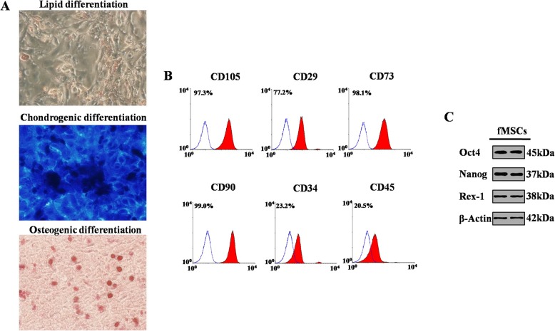
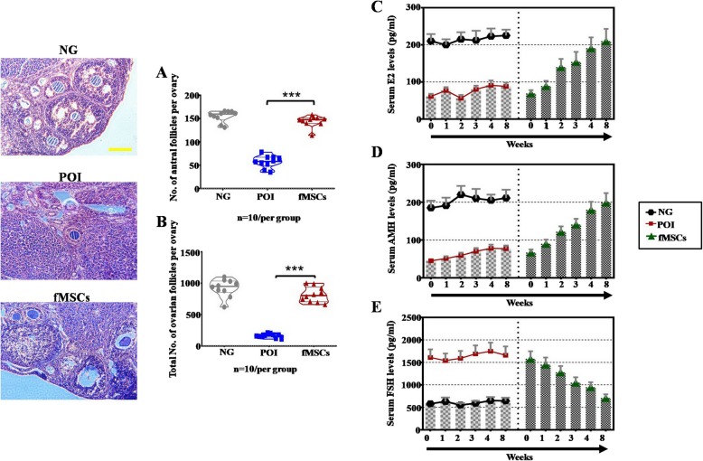
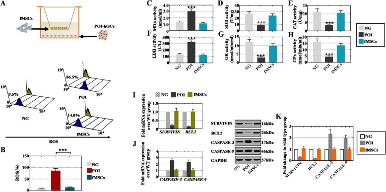
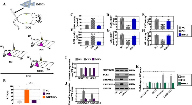
Methods: We designed an in vivo model (chemotherapy-induced ovary damage) and an in vitro model (human ovarian granulosa cells (hGCs)) to understand the efficacy and molecular cues of fMSC treatment of POI. Follicle development was observed by H&E staining. The concentration of sex hormones in serum (E2, AMH, and FSH) and the concentration of oxidative and antioxidative metabolites and the enzymes MDA, SOD, CAT, LDH, GR, and GPx were measured by ELISA. Flow cytometry (FACS) was employed to detect the percentages of ROS and proliferation rates. mRNA and protein expression of antiapoptotic genes (SURVIVIN and BCL2), apoptotic genes (CASPASE-3 and CASPASE-9), and MT1 and its downstream genes (JNK1, PCNA, AMPK) were tested by qPCR and western blotting. MT1 siRNA and related antagonists were used to assess the mechanism.
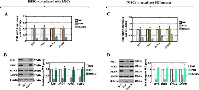
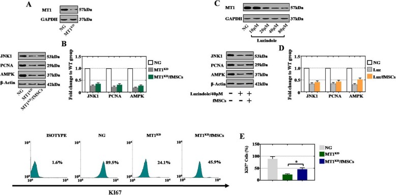
Results: fMSC treatment prevented cyclophosphamide (CTX)-induced follicle loss and recovered sex hormone levels. Additionally, fMSCs significantly decreased oxidative damage, increased oxidative protection, improved antiapoptotic effects, and inhibited apoptotic genes in vivo and in vitro. Furthermore, fMSCs also upregulated MT1, JNK1, PCNA, and AMPK at the mRNA and protein levels. With MT1 knockdown or antagonist treatment in normal hGCs, the protein expression of JNK1, PCNA, and AMPK and the percentage of proliferation were impaired.
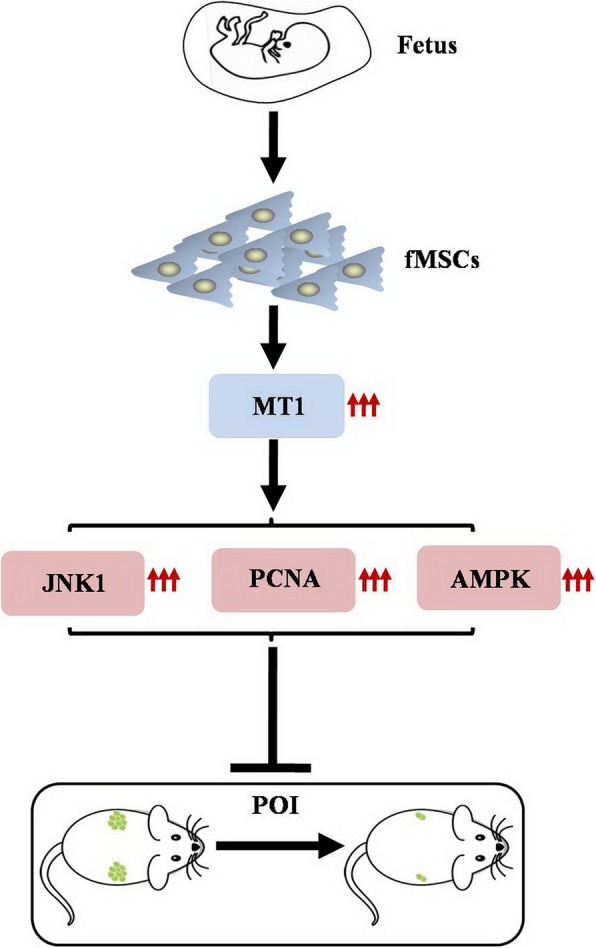
Conclusions: fMSCs might play a crucial role in mediating follicular development in the POI mouse model and stimulating the activity of POI hGCs by targeting MT1.
Keywords: Fetal mesenchymal stem cells; MT1; Premature ovarian insufficiency; Reactive oxygen species.
Conflict of interest statement
The authors declare that they have no competing interests.
Figures
Similar articles
-
Exosomes derived from human adipose mesenchymal stem cells improve ovary function of premature ovarian insufficiency by targeting SMAD.Stem Cell Res Ther. 2018 Aug 9;9(1):216. doi: 10.1186/s13287-018-0953-7. Stem Cell Res Ther. 2018. PMID: 30092819 Free PMC article.
-
Mesenchymal stem cells derived from hPSC via neural crest attenuate chemotherapy-induced premature ovarian insufficiency by ameliorating apoptosis and oxidative stress in granulosa cells.Stem Cell Res Ther. 2025 May 13;16(1):239. doi: 10.1186/s13287-025-04346-x. Stem Cell Res Ther. 2025. PMID: 40361250 Free PMC article.
-
Effects of hPMSCs on granulosa cell apoptosis and AMH expression and their role in the restoration of ovary function in premature ovarian failure mice.Stem Cell Res Ther. 2018 Jan 31;9(1):20. doi: 10.1186/s13287-017-0745-5. Stem Cell Res Ther. 2018. Retraction in: Stem Cell Res Ther. 2022 Oct 13;13(1):504. doi: 10.1186/s13287-022-03183-6. PMID: 29386068 Free PMC article. Retracted.
-
Premature ovarian insufficiency: pathogenesis and therapeutic potential of mesenchymal stem cell.J Mol Med (Berl). 2021 May;99(5):637-650. doi: 10.1007/s00109-021-02055-5. Epub 2021 Feb 27. J Mol Med (Berl). 2021. PMID: 33641066 Review.
-
Human UC-MSC-derived exosomes facilitate ovarian renovation in rats with chemotherapy-induced premature ovarian insufficiency.Front Endocrinol (Lausanne). 2023 Jul 26;14:1205901. doi: 10.3389/fendo.2023.1205901. eCollection 2023. Front Endocrinol (Lausanne). 2023. PMID: 37564988 Free PMC article. Review.
Cited by
-
Vitamin C improves the therapeutic potential of human amniotic epithelial cells in premature ovarian insufficiency disease.Stem Cell Res Ther. 2020 Apr 22;11(1):159. doi: 10.1186/s13287-020-01666-y. Stem Cell Res Ther. 2020. PMID: 32321569 Free PMC article.
-
Therapeutic Effect of Melatonin in Premature Ovarian Insufficiency: Hippo Pathway Is Involved.Oxid Med Cell Longev. 2022 Aug 16;2022:3425877. doi: 10.1155/2022/3425877. eCollection 2022. Oxid Med Cell Longev. 2022. PMID: 36017238 Free PMC article. Review.
-
A Review on Treatment of Premature Ovarian Insufficiency: Characteristics, Limitations, and Challenges of Stem Cell versus ExosomeTherapy.Vet Med Int. 2023 Nov 17;2023:5760011. doi: 10.1155/2023/5760011. eCollection 2023. Vet Med Int. 2023. PMID: 38023426 Free PMC article. Review.
-
Clusterin-carrying extracellular vesicles derived from human umbilical cord mesenchymal stem cells restore the ovarian function of premature ovarian failure mice through activating the PI3K/AKT pathway.Stem Cell Res Ther. 2024 Sep 13;15(1):300. doi: 10.1186/s13287-024-03926-7. Stem Cell Res Ther. 2024. PMID: 39272156 Free PMC article.
-
Application of Mesenchymal Stem Cells in Female Infertility Treatment: Protocols and Preliminary Results.Life (Basel). 2024 Sep 13;14(9):1161. doi: 10.3390/life14091161. Life (Basel). 2024. PMID: 39337944 Free PMC article. Review.
References
-
- Coulam CB, Adamson SC, Annegers JF. Incidence of premature ovarian failure. Obstet Gynecol. 1986;67:604–606. - PubMed
Publication types
MeSH terms
Substances
LinkOut - more resources
Full Text Sources
Medical
Research Materials
Miscellaneous
